8TL4 | pdb_00008tl4

CRYO-EM STRUCTURE OF HIV-1 BG505DS-SOSIP.664 ENV TRIMER BOUND TO DJ85-e.01 FAB


Experimental Data Snapshot

  • Method: ELECTRON MICROSCOPY
  • Resolution: 3.20 Å
  • Aggregation State: PARTICLE 
  • Reconstruction Method: SINGLE PARTICLE 

wwPDB Validation 3D Report Full Report

Validation slider image for 8TL4

This is version 1.2 of the entry. See complete history

Literature

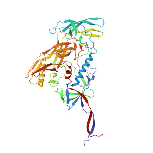
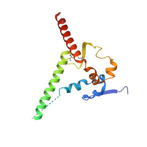
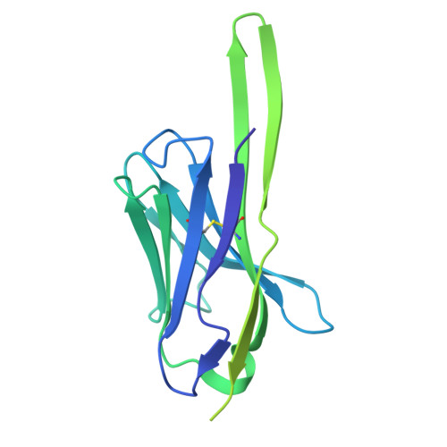
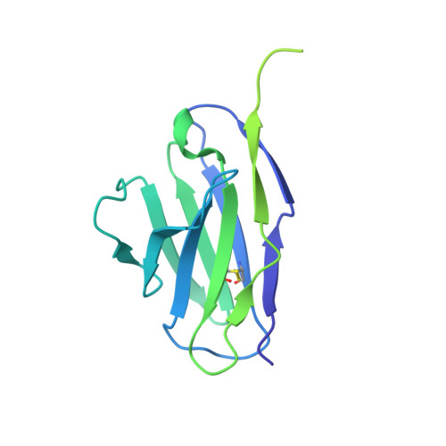
Potent and broad HIV-1 neutralization in fusion peptide-primed SHIV-infected macaques.

Wang, H.Cheng, C.Dal Santo, J.L.Shen, C.H.Bylund, T.Henry, A.R.Howe, C.A.Hwang, J.Morano, N.C.Morris, D.J.Pletnev, S.Roark, R.S.Zhou, T.Hansen, B.T.Hoyt, F.H.Johnston, T.S.Wang, S.Zhang, B.Ambrozak, D.R.Becker, J.E.Bender, M.F.Changela, A.Chaudhary, R.Corcoran, M.Corrigan, A.R.Foulds, K.E.Guo, Y.Lee, M.Li, Y.Lin, B.C.Liu, T.Louder, M.K.Mandolesi, M.Mason, R.D.McKee, K.Nair, V.O'Dell, S.Olia, A.S.Ou, L.Pegu, A.Raju, N.Rawi, R.Roberts-Torres, J.Sarfo, E.K.Sastry, M.Schaub, A.J.Schmidt, S.D.Schramm, C.A.Schwartz, C.L.Smith, S.C.Stephens, T.Stuckey, J.Teng, I.T.Todd, J.P.Tsybovsky, Y.Van Wazer, D.J.Wang, S.Doria-Rose, N.A.Fischer, E.R.Georgiev, I.S.Karlsson Hedestam, G.B.Sheng, Z.Woodward, R.A.Douek, D.C.Koup, R.A.Pierson, T.C.Shapiro, L.Shaw, G.M.Mascola, J.R.Kwong, P.D.

(2024) Cell 187: 7214-7231.e23

  • DOI: https://doi.org/10.1016/j.cell.2024.10.003
  • Primary Citation Related Structures: 
    8TDX, 8TE7, 8TJR, 8TJS, 8TKC, 8TL2, 8TL3, 8TL4, 8TL5, 8TNU, 8TO7, 8TO9, 8TOP

  • PubMed Abstract: 

    An antibody-based HIV-1 vaccine will require the induction of potent cross-reactive HIV-1-neutralizing responses. To demonstrate feasibility toward this goal, we combined vaccination targeting the fusion-peptide site of vulnerability with infection by simian-human immunodeficiency virus (SHIV). In four macaques with vaccine-induced neutralizing responses, SHIV infection boosted plasma neutralization to 45%-77% breadth (geometric mean 50% inhibitory dilution [ID 50 ] ∼100) on a 208-strain panel. Molecular dissection of these responses by antibody isolation and cryo-electron microscopy (cryo-EM) structure determination revealed 15 of 16 antibody lineages with cross-clade neutralization to be directed toward the fusion-peptide site of vulnerability. In each macaque, isolated antibodies from memory B cells recapitulated the plasma-neutralizing response, with fusion-peptide-binding antibodies reaching breadths of 40%-60% (50% inhibitory concentration [IC 50 ] < 50 μg/mL) and total lineage-concentrations estimates of 50-200 μg/mL. Longitudinal mapping indicated that these responses arose prior to SHIV infection. Collectively, these results provide in vivo molecular examples for one to a few B cell lineages affording potent, broadly neutralizing plasma responses.


  • Organizational Affiliation
    • Vaccine Research Center, National Institute of Allergy and Infectious Diseases, National Institutes of Health, Bethesda, MD 20892, USA.

Macromolecule Content 

  • Total Structure Weight: 377.23 kDa 
  • Atom Count: 20,055 
  • Modeled Residue Count: 2,418 
  • Deposited Residue Count: 3,252 
  • Unique protein chains: 4

Macromolecules

Find similar proteins by:|  3D Structure
Entity ID: 1
MoleculeChains  Sequence LengthOrganismDetailsImage
BG505 DS-SOSIP Surface protein gp120
A, C, E
481Human immunodeficiency virus 1Mutation(s): 6 
Gene Names: env
UniProt
Find proteins for Q2N0S6 (Human immunodeficiency virus type 1)
Explore Q2N0S6 
Go to UniProtKB:  Q2N0S6
Entity Groups
Sequence Clusters30% Identity50% Identity70% Identity90% Identity95% Identity100% Identity
UniProt GroupQ2N0S6
Glycosylation
Glycosylation Sites: 15
Sequence Annotations
Expand
Reference Sequence
Find similar proteins by:|  3D Structure
Entity ID: 2
MoleculeChains  Sequence LengthOrganismDetailsImage
BG505 DS-SOSIP Transmembrane protein gp41
B, D, F
153Human immunodeficiency virus 1Mutation(s): 2 
Gene Names: env
UniProt
Find proteins for Q2N0S5 (Human immunodeficiency virus type 1)
Explore Q2N0S5 
Go to UniProtKB:  Q2N0S5
Entity Groups
Sequence Clusters30% Identity50% Identity70% Identity90% Identity95% Identity100% Identity
UniProt GroupQ2N0S5
Sequence Annotations
Expand
Reference Sequence
Find similar proteins by:|  3D Structure
Entity ID: 3
MoleculeChains  Sequence LengthOrganismDetailsImage
DJ85-e.01 FAB HEAVY CHAIN
G, I, K
236Homo sapiensMutation(s): 0 
Find similar proteins by:|  3D Structure
Entity ID: 4
MoleculeChains  Sequence LengthOrganismDetailsImage
DJ85-e.01 FAB LIGHT CHAIN
H, J, L
214Homo sapiensMutation(s): 0 

Oligosaccharides

Help  
Entity ID: 5
MoleculeChains Length2D Diagram GlycosylationD Interactions
2-acetamido-2-deoxy-beta-D-glucopyranose-(1-4)-2-acetamido-2-deoxy-beta-D-glucopyranose
BA [auth Z],
CA [auth b],
DA [auth d],
EA [auth e],
FA [auth f],
BA [auth Z],
CA [auth b],
DA [auth d],
EA [auth e],
FA [auth f],
GA [auth g],
HA [auth h],
IA [auth i],
KA [auth k],
LA [auth l],
M [auth a],
MA [auth m],
N [auth M],
O [auth N],
P [auth O],
Q [auth P],
S [auth R],
T [auth S],
U [auth T],
V [auth c],
W [auth U],
X [auth V],
Y [auth W],
Z [auth X]
2N-Glycosylation
Glycosylation Resources
GlyTouCan: G42666HT
GlyCosmos: G42666HT
GlyGen: G42666HT
Entity ID: 6
MoleculeChains Length2D Diagram GlycosylationD Interactions
alpha-D-mannopyranose-(1-2)-alpha-D-mannopyranose-(1-3)-beta-D-mannopyranose-(1-4)-2-acetamido-2-deoxy-beta-D-glucopyranose-(1-4)-2-acetamido-2-deoxy-beta-D-glucopyranoseAA [auth Y],
JA [auth j],
R [auth Q]
5N-Glycosylation
Glycosylation Resources
GlyTouCan: G42227JK
GlyCosmos: G42227JK
GlyGen: G42227JK

Small Molecules

Ligands 1 Unique
IDChains Name / Formula / InChI Key2D Diagram3D Interactions
NAG

Query on NAG


Download:Ideal Coordinates CCD File
AB [auth E]
BB [auth E]
CB [auth E]
DB [auth E]
EB [auth E]
AB [auth E],
BB [auth E],
CB [auth E],
DB [auth E],
EB [auth E],
NA [auth A],
OA [auth A],
PA [auth A],
QA [auth A],
RA [auth A],
SA [auth A],
TA [auth C],
UA [auth C],
VA [auth C],
WA [auth C],
XA [auth C],
YA [auth C],
ZA [auth E]
2-acetamido-2-deoxy-beta-D-glucopyranose
C8 H15 N O6
OVRNDRQMDRJTHS-FMDGEEDCSA-N

Experimental Data & Validation

Experimental Data

  • Method: ELECTRON MICROSCOPY
  • Resolution: 3.20 Å
  • Aggregation State: PARTICLE 
  • Reconstruction Method: SINGLE PARTICLE 
EM Software:
TaskSoftware PackageVersion
MODEL REFINEMENTPHENIX1.20

Structure Validation

View Full Validation Report


Entry History 

& Funding Information

Deposition Data


Funding OrganizationLocationGrant Number
National Institutes of Health/National Institute Of Allergy and Infectious Diseases (NIH/NIAID)United States--

Revision History  (Full details and data files)

  • Version 1.0: 2024-08-28
    Type: Initial release
  • Version 1.1: 2024-10-23
    Changes: Data collection, Structure summary
  • Version 1.2: 2025-01-15
    Changes: Data collection, Database references