8TK8

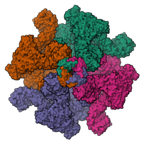
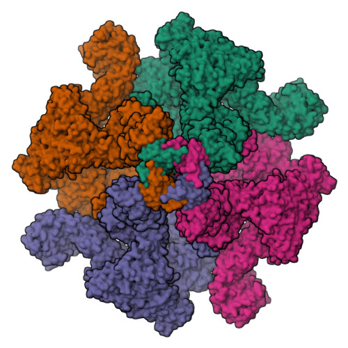
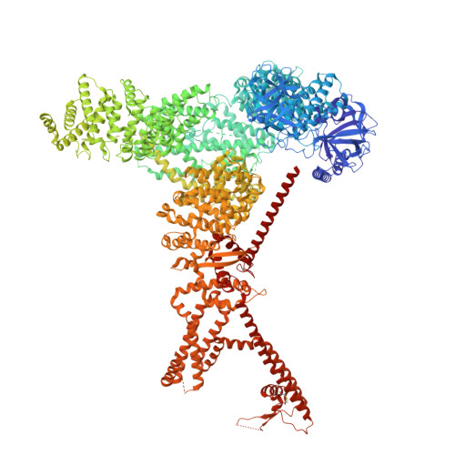
Human Type 3 IP3 Receptor - Resting State (+IP3/ATP)

  • Classification: TRANSPORT PROTEIN
  • Organism(s): Homo sapiens
  • Expression System: Homo sapiens
  • Mutation(s): No 
  • Membrane Protein: Yes  mpstruc

  • Deposited: 2023-07-25 Released: 2023-11-08 
  • Deposition Author(s): Paknejad, N., Sapuru, V., Hite, R.K.
  • Funding Organization(s): National Institutes of Health/National Institute of General Medical Sciences (NIH/NIGMS), National Institutes of Health/National Cancer Institute (NIH/NCI)

Experimental Data Snapshot

  • Method: ELECTRON MICROSCOPY
  • Resolution: 2.70 Å
  • Aggregation State: PARTICLE 
  • Reconstruction Method: SINGLE PARTICLE 

wwPDB Validation   3D Report Full Report


This is version 1.0 of the entry. See complete history


Literature

Structural titration reveals Ca 2+ -dependent conformational landscape of the IP 3 receptor.

Paknejad, N.Sapuru, V.Hite, R.K.

(2023) Nat Commun 14: 6897-6897

  • DOI: https://doi.org/10.1038/s41467-023-42707-3
  • Primary Citation of Related Structures:  
    8TK8, 8TKD, 8TKE, 8TKF, 8TKG, 8TKH, 8TKI, 8TL9, 8TLA

  • PubMed Abstract: 

    Inositol 1,4,5-trisphosphate receptors (IP 3 Rs) are endoplasmic reticulum Ca 2+ channels whose biphasic dependence on cytosolic Ca 2+ gives rise to Ca 2+ oscillations that regulate fertilization, cell division and cell death. Despite the critical roles of IP 3 R-mediated Ca 2+ responses, the structural underpinnings of the biphasic Ca 2+ dependence that underlies Ca 2+ oscillations are incompletely understood. Here, we collect cryo-EM images of an IP 3 R with Ca 2+ concentrations spanning five orders of magnitude. Unbiased image analysis reveals that Ca 2+ binding does not explicitly induce conformational changes but rather biases a complex conformational landscape consisting of resting, preactivated, activated, and inhibited states. Using particle counts as a proxy for relative conformational free energy, we demonstrate that Ca 2+ binding at a high-affinity site allows IP 3 Rs to activate by escaping a low-energy resting state through an ensemble of preactivated states. At high Ca 2+ concentrations, IP 3 Rs preferentially enter an inhibited state stabilized by a second, low-affinity Ca 2+ binding site. Together, these studies provide a mechanistic basis for the biphasic Ca 2+ -dependence of IP 3 R channel activity.


  • Organizational Affiliation

    Structural Biology Program, Memorial Sloan Kettering Cancer Center, New York, NY, 10065, USA.


Macromolecules
Find similar proteins by:  (by identity cutoff)  |  3D Structure
Entity ID: 1
MoleculeChains Sequence LengthOrganismDetailsImage
Inositol 1,4,5-trisphosphate receptor type 3
A, B, C, D
2,671Homo sapiensMutation(s): 0 
Gene Names: ITPR3
Membrane Entity: Yes 
UniProt & NIH Common Fund Data Resources
Find proteins for Q14573 (Homo sapiens)
Explore Q14573 
Go to UniProtKB:  Q14573
PHAROS:  Q14573
GTEx:  ENSG00000096433 
Entity Groups  
Sequence Clusters30% Identity50% Identity70% Identity90% Identity95% Identity100% Identity
UniProt GroupQ14573
Sequence Annotations
Expand
  • Reference Sequence
Small Molecules
Ligands 4 Unique
IDChains Name / Formula / InChI Key2D Diagram3D Interactions
PCW
Query on PCW

Download Ideal Coordinates CCD File 
AA [auth C]
BA [auth C]
CA [auth C]
DA [auth C]
EA [auth C]
AA [auth C],
BA [auth C],
CA [auth C],
DA [auth C],
EA [auth C],
FA [auth C],
GA [auth D],
H [auth A],
HA [auth D],
I [auth A],
J [auth A],
K [auth A],
L [auth A],
LA [auth D],
M [auth A],
MA [auth D],
N [auth A],
NA [auth D],
O [auth B],
S [auth B],
T [auth B],
U [auth B],
V [auth B],
W [auth C]
1,2-DIOLEOYL-SN-GLYCERO-3-PHOSPHOCHOLINE
C44 H85 N O8 P
SNKAWJBJQDLSFF-NVKMUCNASA-O
ATP (Subject of Investigation/LOI)
Query on ATP

Download Ideal Coordinates CCD File 
G [auth A],
KA [auth D],
R [auth B],
Z [auth C]
ADENOSINE-5'-TRIPHOSPHATE
C10 H16 N5 O13 P3
ZKHQWZAMYRWXGA-KQYNXXCUSA-N
I3P (Subject of Investigation/LOI)
Query on I3P

Download Ideal Coordinates CCD File 
F [auth A],
JA [auth D],
Q [auth B],
Y [auth C]
D-MYO-INOSITOL-1,4,5-TRIPHOSPHATE
C6 H15 O15 P3
MMWCIQZXVOZEGG-XJTPDSDZSA-N
ZN (Subject of Investigation/LOI)
Query on ZN

Download Ideal Coordinates CCD File 
E [auth A],
IA [auth D],
P [auth B],
X [auth C]
ZINC ION
Zn
PTFCDOFLOPIGGS-UHFFFAOYSA-N
Binding Affinity Annotations 
IDSourceBinding Affinity
I3P BindingDB:  8TK8 Kd: 195 (nM) from 1 assay(s)
IC50: 1.40e+4 (nM) from 1 assay(s)
Experimental Data & Validation

Experimental Data

  • Method: ELECTRON MICROSCOPY
  • Resolution: 2.70 Å
  • Aggregation State: PARTICLE 
  • Reconstruction Method: SINGLE PARTICLE 
EM Software:
TaskSoftware PackageVersion
RECONSTRUCTIONcryoSPARC
MODEL REFINEMENTPHENIXdev_4761:

Structure Validation

View Full Validation Report


Entry History & Funding Information

Deposition Data


Funding OrganizationLocationGrant Number
National Institutes of Health/National Institute of General Medical Sciences (NIH/NIGMS)United StatesGM13230704
National Institutes of Health/National Cancer Institute (NIH/NCI)United StatesCA008748
National Institutes of Health/National Cancer Institute (NIH/NCI)United StatesCA243235

Revision History  (Full details and data files)

  • Version 1.0: 2023-11-08
    Type: Initial release