8T1X | pdb_00008t1x

T4 highly immunogenic outer capsid protein C-terminal domain bound to the vertex-proximal gp23* capsomer of the isometric head in two preferred orientations

  • Classification: VIRAL PROTEIN
  • Organism(s): Escherichia phage T4
  • Mutation(s): Yes 

  • Deposited: 2023-06-04 Released: 2023-07-19 
  • Deposition Author(s): Fokine, A., Rao, V.B.
  • Funding Organization(s): National Institutes of Health/National Institute Of Allergy and Infectious Diseases (NIH/NIAID)

Experimental Data Snapshot

  • Method: ELECTRON MICROSCOPY
  • Resolution: 3.30 Å
  • Aggregation State: PARTICLE 
  • Reconstruction Method: SINGLE PARTICLE 

Starting Model: in silico
View more details

wwPDB Validation 3D Report Full Report

Validation slider image for 8T1X

This is version 1.3 of the entry. See complete history

Literature

Structure and Function of Hoc-A Novel Environment Sensing Device Encoded by T4 and Other Bacteriophages.

Fokine, A.Islam, M.Z.Fang, Q.Chen, Z.Sun, L.Rao, V.B.

(2023) Viruses 15

  • DOI: https://doi.org/10.3390/v15071517
  • Primary Citation Related Structures: 
    8T1X, 8T9R

  • PubMed Abstract: 

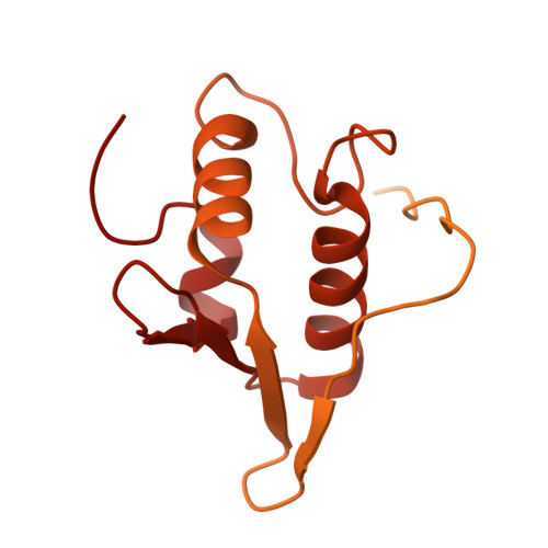
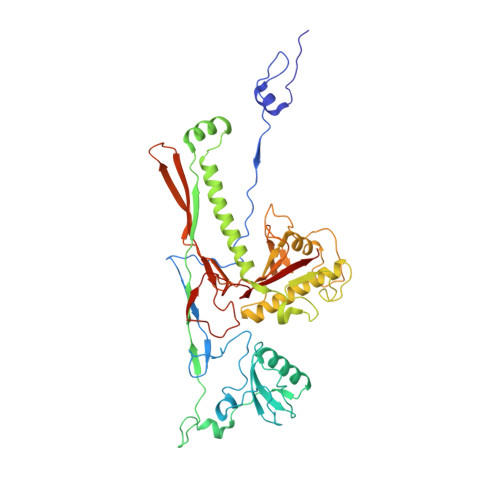
    Bacteriophage T4 is decorated with 155 180 Å-long fibers of the highly antigenic outer capsid protein (Hoc). In this study, we describe a near-atomic structural model of Hoc by combining cryo-electron microscopy and AlphaFold structure predictions. It consists of a conserved C-terminal capsid-binding domain attached to a string of three variable immunoglobulin (Ig)-like domains, an architecture well-preserved in hundreds of Hoc molecules found in phage genomes. Each T4-Hoc fiber attaches randomly to the center of gp23* hexameric capsomers in one of the six possible orientations, though at the vertex-proximal hexamers that deviate from 6-fold symmetry, Hoc binds in two preferred orientations related by 180° rotation. Remarkably, each Hoc fiber binds to all six subunits of the capsomer, though the interactions are greatest with three of the subunits, resulting in the off-centered attachment of the C-domain. Biochemical analyses suggest that the acidic Hoc fiber (pI, ~4-5) allows for the clustering of virions in acidic pH and dispersion in neutral/alkaline pH. Hoc appears to have evolved as a sensing device that allows the phage to navigate its movements through reversible clustering-dispersion transitions so that it reaches its destination, the host bacterium, and persists in various ecological niches such as the human/mammalian gut.


  • Organizational Affiliation
    • Department of Biological Sciences, Purdue University, West Lafayette, IN 47907, USA.

Macromolecule Content 

  • Total Structure Weight: 373.21 kDa 
  • Atom Count: 22,172 
  • Modeled Residue Count: 2,928 
  • Deposited Residue Count: 3,488 
  • Unique protein chains: 2

Macromolecules

Find similar proteins by:|  3D Structure
Entity ID: 1
MoleculeChains  Sequence LengthOrganismDetailsImage
Highly immunogenic outer capsid proteinA [auth X],
B [auth Y]
376Escherichia phage T4Mutation(s): 0 
UniProt
Find proteins for P18056 (Enterobacteria phage T4)
Explore P18056 
Go to UniProtKB:  P18056
Entity Groups
Sequence Clusters30% Identity50% Identity70% Identity90% Identity95% Identity100% Identity
UniProt GroupP18056
Sequence Annotations
Expand
Reference Sequence
Find similar proteins by:|  3D Structure
Entity ID: 2
MoleculeChains  Sequence LengthOrganismDetailsImage
Mature major capsid protein gp23*456Escherichia phage T4Mutation(s): 1 
UniProt
Find proteins for P04535 (Enterobacteria phage T4)
Explore P04535 
Go to UniProtKB:  P04535
Entity Groups
Sequence Clusters30% Identity50% Identity70% Identity90% Identity95% Identity100% Identity
UniProt GroupP04535
Sequence Annotations
Expand
Reference Sequence

Experimental Data & Validation

Experimental Data

  • Method: ELECTRON MICROSCOPY
  • Resolution: 3.30 Å
  • Aggregation State: PARTICLE 
  • Reconstruction Method: SINGLE PARTICLE 
EM Software:
TaskSoftware PackageVersion
MODEL REFINEMENTPHENIX

Structure Validation

View Full Validation Report


Entry History 

& Funding Information

Deposition Data


Funding OrganizationLocationGrant Number
National Institutes of Health/National Institute Of Allergy and Infectious Diseases (NIH/NIAID)United States--

Revision History  (Full details and data files)

  • Version 1.0: 2023-07-19
    Type: Initial release
  • Version 1.1: 2023-08-09
    Changes: Database references
  • Version 1.2: 2024-06-19
    Changes: Data collection
  • Version 1.3: 2026-03-04
    Changes: Refinement description, Structure summary