8SJK

Pembrolizumab Caffeine crystal


Experimental Data Snapshot

  • Method: X-RAY DIFFRACTION
  • Resolution: 2.22 Å
  • R-Value Free: 0.238 
  • R-Value Work: 0.186 
  • R-Value Observed: 0.189 

Starting Model: experimental
View more details

wwPDB Validation   3D Report Full Report


Ligand Structure Quality Assessment 


This is version 1.1 of the entry. See complete history


Literature

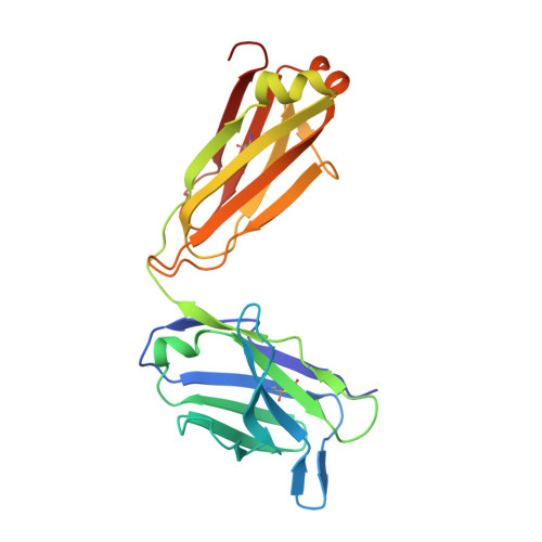
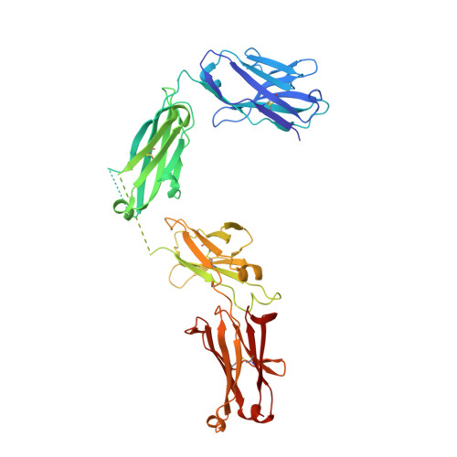
Small-Angle X-ray Scattering as a Powerful Tool for Phase and Crystallinity Assessment of Monoclonal Antibody Crystallites in Support of Batch Crystallization.

Larpent, P.Codan, L.Bothe, J.R.Iuzzolino, L.Pabit, S.Gupta, S.Fischmann, T.Su, Y.Reichert, P.Stueber, D.Cote, A.

(2024) Mol Pharm 

  • DOI: https://doi.org/10.1021/acs.molpharmaceut.4c00418
  • Primary Citation of Related Structures:  
    8SJK

  • PubMed Abstract: 

    Crystalline suspensions of monoclonal antibodies (mAbs) have great potential to improve drug substance isolation and purification on a large scale and to be used for drug delivery via high-concentration formulations. Crystalline mAb suspensions are expected to have enhanced chemical and physical properties relative to mAb solutions delivered intravenously, making them attractive candidates for subcutaneous delivery. In contrast to small molecules, the development of protein crystalline suspensions is not a widely used approach in the pharmaceutical industry. This is mainly due to the challenges in finding crystalline hits and the suboptimal physical properties of the resulting crystallites when hits are found. Modern advances in instrumentation and increased knowledge of mAb crystallization have, however, resulted in higher probabilities of discovering crystal forms and improving their particle properties and characterization. In this regard, physical, analytical characterization plays a central role in the initial steps of understanding and later optimizing the crystallization of mAbs and requires careful selection of the appropriate tools. This contribution describes a novel crystal structure of the antibody pembrolizumab and demonstrates the usefulness of small-angle X-ray scattering (SAXS) for characterizing its crystalline suspensions. It illustrates the advantages of SAXS when used to (i) confirm crystallinity and crystal phase of crystallites produced in batch mode; (ii) confirm crystallinity under various conditions and detect variations in crystal phases, enabling fine-tuning of the crystallizations for phase control across multiple batches; (iii) monitor the physical response and stability of the crystallites in suspension with regard to filtration and washing; and (iv) monitor the physical stability of the crystallites upon drying. Overall, this work highlights how SAXS is an essential tool for mAb crystallization characterization.


  • Organizational Affiliation

    Department of Analytical Research and Development, MSD Werthenstein BioPharma GmbH, Industrie Nord 1, 6105 Schachen, Switzerland.


Macromolecules
Find similar proteins by:  (by identity cutoff)  |  3D Structure
Entity ID: 1
MoleculeChains Sequence LengthOrganismDetailsImage
ANTIBODY LIGHT CHAIN218Homo sapiensMutation(s): 0 
Entity Groups  
Sequence Clusters30% Identity50% Identity70% Identity90% Identity95% Identity100% Identity
Sequence Annotations
Expand
  • Reference Sequence
Find similar proteins by:  (by identity cutoff)  |  3D Structure
Entity ID: 2
MoleculeChains Sequence LengthOrganismDetailsImage
ANTIBODY HEAVY CHAIN444Homo sapiensMutation(s): 0 
Entity Groups  
Sequence Clusters30% Identity50% Identity70% Identity90% Identity95% Identity100% Identity
Glycosylation
Glycosylation Sites: 1
Sequence Annotations
Expand
  • Reference Sequence
Oligosaccharides

Help

Entity ID: 3
MoleculeChains Length2D Diagram Glycosylation3D Interactions
2-acetamido-2-deoxy-beta-D-glucopyranose-(1-2)-alpha-D-mannopyranose-(1-3)-[2-acetamido-2-deoxy-beta-D-glucopyranose-(1-2)-alpha-D-mannopyranose-(1-6)]beta-D-mannopyranose-(1-4)-2-acetamido-2-deoxy-beta-D-glucopyranose-(1-4)-2-acetamido-2-deoxy-beta-D-glucopyranoseC [auth D]7N-Glycosylation
Glycosylation Resources
GlyTouCan:  G39213VZ
GlyCosmos:  G39213VZ
GlyGen:  G39213VZ
Entity ID: 4
MoleculeChains Length2D Diagram Glycosylation3D Interactions
beta-D-fructofuranose-(2-1)-alpha-D-glucopyranoseD [auth E],
E [auth F]
2N/A
Glycosylation Resources
GlyTouCan:  G05551OP
GlyCosmos:  G05551OP
Small Molecules
Ligands 1 Unique
IDChains Name / Formula / InChI Key2D Diagram3D Interactions
CFF (Subject of Investigation/LOI)
Query on CFF

Download Ideal Coordinates CCD File 
F [auth A]CAFFEINE
C8 H10 N4 O2
RYYVLZVUVIJVGH-UHFFFAOYSA-N
Biologically Interesting Molecules (External Reference) 1 Unique
Experimental Data & Validation

Experimental Data

  • Method: X-RAY DIFFRACTION
  • Resolution: 2.22 Å
  • R-Value Free: 0.238 
  • R-Value Work: 0.186 
  • R-Value Observed: 0.189 
  • Space Group: P 2 2 21
Unit Cell:
Length ( Å )Angle ( ˚ )
a = 43.8α = 90
b = 113.9β = 90
c = 175γ = 90
Software Package:
Software NamePurpose
BUSTERrefinement
XDSdata reduction
MOLREPphasing
Aimlessdata scaling

Structure Validation

View Full Validation Report


Ligand Structure Quality Assessment 


Entry History & Funding Information

Deposition Data


Funding OrganizationLocationGrant Number
Other privateUnited States--

Revision History  (Full details and data files)

  • Version 1.0: 2024-04-24
    Type: Initial release
  • Version 1.1: 2024-07-17
    Changes: Database references