


Crystal structure of MBP fusion with HPPK from Methanocaldococcus jannaschii
Shaw, G.X., Needle, D., Stair, N.R., Cherry, S., Tropea, J.E., Waugh, D.S., Ji, X.To be published.
Experimental Data Snapshot
Starting Model: experimental
View more details
wwPDB Validation 3D Report Full Report

Entity ID: 1 | |||||
|---|---|---|---|---|---|
| Molecule | Chains | Sequence Length | Organism | Details | Image |
| Maltose/maltodextrin-binding periplasmic protein,6-hydroxymethyl-7,8-dihydropterin pyrophosphokinase | 618 | Escherichia coli K-12, Methanocaldococcus jannaschii DSM 2661 This entity is chimeric | Mutation(s): 12 Gene Names: malE, b4034, JW3994, mptE, MJ1634 EC: 2.7.6.3 | 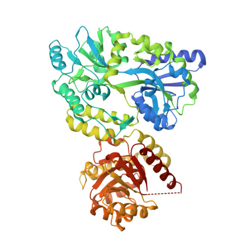 | |
UniProt | |||||
Entity Groups | |||||
| Sequence Clusters | 30% Identity50% Identity70% Identity90% Identity95% Identity100% Identity | ||||
| UniProt Groups | P0AEX9Q59028 | ||||
Sequence AnnotationsExpand | |||||
Reference Sequence | |||||
Entity ID: 2 | |||||
|---|---|---|---|---|---|
| ID | Chains | Name | Type/Class | 2D Diagram | 3D Interactions |
| PRD_900001 Query on PRD_900001 | C, D | alpha-maltose | Oligosaccharide / Nutrient |  |
| Length ( Å ) | Angle ( ˚ ) |
|---|---|
| a = 58.17 | α = 67.44 |
| b = 69.827 | β = 74.63 |
| c = 73.121 | γ = 78.13 |
| Software Name | Purpose |
|---|---|
| PHENIX | refinement |
| SCALEPACK | data scaling |
| DENZO | data reduction |
| PHASER | phasing |
| Funding Organization | Location | Grant Number |
|---|---|---|
| National Institutes of Health/National Cancer Institute (NIH/NCI) | United States | Intramural Research Program |


