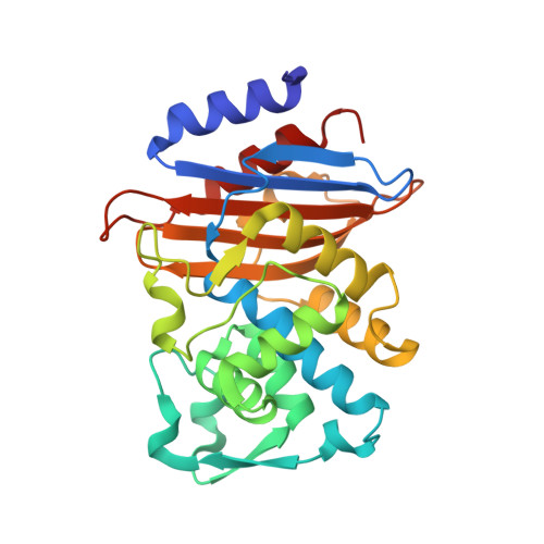
8R7M

CTX-M14 in complex with boric acid and 1,2-diol boric ester


Experimental Data Snapshot

  • Method: X-RAY DIFFRACTION
  • Resolution: 1.00 Å
  • R-Value Free: 0.138 
  • R-Value Work: 0.129 
  • R-Value Observed: 0.129 

wwPDB Validation   3D Report Full Report


Ligand Structure Quality Assessment 


This is version 1.1 of the entry. See complete history


Literature

Time-resolved crystallography of boric acid binding to the active site serine of the beta-lactamase CTX-M-14 and subsequent 1,2-diol esterification.

Prester, A.Perbandt, M.Galchenkova, M.Oberthuer, D.Werner, N.Henkel, A.Maracke, J.Yefanov, O.Hakanpaa, J.Pompidor, G.Meyer, J.Chapman, H.Aepfelbacher, M.Hinrichs, W.Rohde, H.Betzel, C.

(2024) Commun Chem 7: 152-152

  • DOI: https://doi.org/10.1038/s42004-024-01236-w
  • Primary Citation of Related Structures:  
    8PC9, 8PCA, 8PCB, 8PCC, 8PCD, 8PCE, 8PCF, 8PCG, 8PCI, 8PCJ, 8PCK, 8PCL, 8PCM, 8PCN, 8PCO, 8PCP, 8PCQ, 8PCR, 8PCS, 8PCT, 8PCU, 8PCV, 8R7M

  • PubMed Abstract: 

    The emergence and spread of antibiotic resistance represent a growing threat to public health. Of particular concern is the appearance of β-lactamases, which are capable to hydrolyze and inactivate the most important class of antibiotics, the β-lactams. Effective β-lactamase inhibitors and mechanistic insights into their action are central in overcoming this type of resistance, and in this context boronate-based β-lactamase inhibitors were just recently approved to treat multidrug-resistant bacteria. Using boric acid as a simplified inhibitor model, time-resolved serial crystallography was employed to obtain mechanistic insights into binding to the active site serine of β-lactamase CTX-M-14, identifying a reaction time frame of 80-100 ms. In a next step, the subsequent 1,2-diol boric ester formation with glycerol in the active site was monitored proceeding in a time frame of 100-150 ms. Furthermore, the displacement of the crucial anion in the active site of the β-lactamase was verified as an essential part of the binding mechanism of substrates and inhibitors. In total, 22 datasets of β-lactamase intermediate complexes with high spatial resolution of 1.40-2.04 Å and high temporal resolution range of 50-10,000 ms were obtained, allowing a detailed analysis of the studied processes. Mechanistic details captured here contribute to the understanding of molecular processes and their time frames in enzymatic reactions. Moreover, we could demonstrate that time-resolved crystallography can serve as an additional tool for identifying and investigating enzymatic reactions.


  • Organizational Affiliation

    Institute of Medical Microbiology, Virology and Hygiene, University Medical Center Hamburg-Eppendorf UKE, Hamburg, Germany.


Macromolecules
Find similar proteins by:  (by identity cutoff)  |  3D Structure
Entity ID: 1
MoleculeChains Sequence LengthOrganismDetailsImage
Beta-lactamase263Klebsiella pneumoniaeMutation(s): 0 
EC: 3.5.2.6
UniProt
Find proteins for A0A2S1JJX2 (Klebsiella pneumoniae)
Explore A0A2S1JJX2 
Go to UniProtKB:  A0A2S1JJX2
Entity Groups  
Sequence Clusters30% Identity50% Identity70% Identity90% Identity95% Identity100% Identity
UniProt GroupA0A2S1JJX2
Sequence Annotations
Expand
  • Reference Sequence
Small Molecules
Ligands 5 Unique
IDChains Name / Formula / InChI Key2D Diagram3D Interactions
YCH (Subject of Investigation/LOI)
Query on YCH

Download Ideal Coordinates CCD File 
D [auth A][(4~{S})-2-oxidanyl-1,3,2-dioxaborolan-4-yl]methanol
C3 H7 B O4
HXGHIZVNPXUHFK-VKHMYHEASA-N
SO4
Query on SO4

Download Ideal Coordinates CCD File 
G [auth A],
H [auth A]
SULFATE ION
O4 S
QAOWNCQODCNURD-UHFFFAOYSA-L
GOL
Query on GOL

Download Ideal Coordinates CCD File 
C [auth A],
E [auth A],
F [auth A]
GLYCEROL
C3 H8 O3
PEDCQBHIVMGVHV-UHFFFAOYSA-N
BO3 (Subject of Investigation/LOI)
Query on BO3

Download Ideal Coordinates CCD File 
B [auth A]BORIC ACID
B H3 O3
KGBXLFKZBHKPEV-UHFFFAOYSA-N
NA
Query on NA

Download Ideal Coordinates CCD File 
I [auth A]SODIUM ION
Na
FKNQFGJONOIPTF-UHFFFAOYSA-N
Experimental Data & Validation

Experimental Data

  • Method: X-RAY DIFFRACTION
  • Resolution: 1.00 Å
  • R-Value Free: 0.138 
  • R-Value Work: 0.129 
  • R-Value Observed: 0.129 
  • Space Group: P 21 21 21
Unit Cell:
Length ( Å )Angle ( ˚ )
a = 41.775α = 90
b = 62.104β = 90
c = 86.508γ = 90
Software Package:
Software NamePurpose
PHENIXrefinement
XDSdata reduction
XDSdata scaling
PHASERphasing

Structure Validation

View Full Validation Report


Ligand Structure Quality Assessment 


Entry History & Funding Information

Deposition Data


Funding OrganizationLocationGrant Number
German Federal Ministry for Education and ResearchGermany05K13GUA

Revision History  (Full details and data files)

  • Version 1.0: 2024-01-17
    Type: Initial release
  • Version 1.1: 2024-07-17
    Changes: Database references