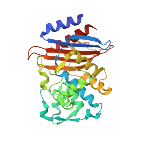
Time-resolved crystallography of boric acid binding to the active site serine of the beta-lactamase CTX-M-14 and subsequent 1,2-diol esterification.
Prester, A., Perbandt, M., Galchenkova, M., Oberthuer, D., Werner, N., Henkel, A., Maracke, J., Yefanov, O., Hakanpaa, J., Pompidor, G., Meyer, J., Chapman, H., Aepfelbacher, M., Hinrichs, W., Rohde, H., Betzel, C.(2024) Commun Chem 7: 152-152
- PubMed: 38969718 Search on PubMedSearch on PubMed Central
- DOI: https://doi.org/10.1038/s42004-024-01236-w
- Primary Citation Related Structures:
8PC9, 8PCA, 8PCB, 8PCC, 8PCD, 8PCE, 8PCF, 8PCG, 8PCI, 8PCJ, 8PCK, 8PCL, 8PCM, 8PCN, 8PCO, 8PCP, 8PCQ, 8PCR, 8PCS, 8PCT, 8PCU, 8PCV, 8R7M - PubMed Abstract:
The emergence and spread of antibiotic resistance represent a growing threat to public health. Of particular concern is the appearance of β-lactamases, which are capable to hydrolyze and inactivate the most important class of antibiotics, the β-lactams. Effective β-lactamase inhibitors and mechanistic insights into their action are central in overcoming this type of resistance, and in this context boronate-based β-lactamase inhibitors were just recently approved to treat multidrug-resistant bacteria. Using boric acid as a simplified inhibitor model, time-resolved serial crystallography was employed to obtain mechanistic insights into binding to the active site serine of β-lactamase CTX-M-14, identifying a reaction time frame of 80-100 ms. In a next step, the subsequent 1,2-diol boric ester formation with glycerol in the active site was monitored proceeding in a time frame of 100-150 ms. Furthermore, the displacement of the crucial anion in the active site of the β-lactamase was verified as an essential part of the binding mechanism of substrates and inhibitors. In total, 22 datasets of β-lactamase intermediate complexes with high spatial resolution of 1.40-2.04 Å and high temporal resolution range of 50-10,000 ms were obtained, allowing a detailed analysis of the studied processes. Mechanistic details captured here contribute to the understanding of molecular processes and their time frames in enzymatic reactions. Moreover, we could demonstrate that time-resolved crystallography can serve as an additional tool for identifying and investigating enzymatic reactions.
- Institute of Medical Microbiology, Virology and Hygiene, University Medical Center Hamburg-Eppendorf UKE, Hamburg, Germany.
Organizational Affiliation: