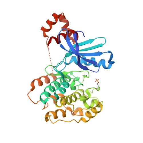
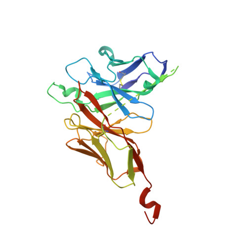
8R4O

Salt inducible kinase 3 in complex with inhibitor


Experimental Data Snapshot

  • Method: X-RAY DIFFRACTION
  • Resolution: 2.73 Å
  • R-Value Free: 0.244 
  • R-Value Work: 0.214 
  • R-Value Observed: 0.216 

wwPDB Validation   3D Report Full Report


Ligand Structure Quality Assessment 


This is version 1.1 of the entry. See complete history


Literature

The structures of salt inducible kinase 3 in complex with pharmacological inhibitors reveal determinants for binding and selectivity.

Oster, L.Castaldo, M.de Vries, E.Edfeldt, F.Pemberton, N.Gordon, E.Cederblad, L.Kack, H.

(2024) J Biol Chem : 107201-107201

  • DOI: https://doi.org/10.1016/j.jbc.2024.107201
  • Primary Citation of Related Structures:  
    8R4O, 8R4Q, 8R4U, 8R4V

  • PubMed Abstract: 

    The salt-inducible kinases (SIKs) 1-3, belonging to the AMPK-related kinase family, serve as master regulators orchestrating a diverse set of physiological processes such as metabolism, bone formation, immune response, oncogenesis and cardiac rhythm. Owing to its key regulatory role, the SIK kinases have emerged as compelling targets for pharmacological intervention across a diverse set of indications. Therefore, there is interest in developing SIK inhibitors with defined selectivity profiles both to further dissect the downstream biology and for treating disease. However, despite a large pharmaceutical interest in the SIKs, experimental structures of SIK kinases are scarce. This is likely due to the challenges associated with generation of protein suitable for structural studies. By adopting a rational approach to construct design and protein purification we successfully crystallized and subsequently solved the structure of SIK3 in complex with HG-9-91-01, a potent SIK inhibitor. To enable further SIK3-inhibitor complex structures we identified an antibody fragment which facilitated crystallization and enabled a robust protocol suitable for structure-based drug-design. The structures reveal SIK3 in an active conformation, where the ubiquitin-associated domain is shown to provide further stabilization to this active conformation. We present four pharmacologically relevant and distinct SIK3-inhibitor complexes. These detail the key interaction for each ligand and reveal how different regions of the ATP site are engaged by the different inhibitors to achieve high affinity. Notably, the structure of SIK3 in complex with a SIK3 specific inhibitor, offers insights into isoform selectivity.


  • Organizational Affiliation

    Mechanistic and Structural Biology, Discovery Sciences, BioPharmaceuticals R&D, AstraZeneca, Pepparedsleden 1, 43183, Gothenburg, Sweden. Electronic address: linda.oster@astrazeneca.com.


Macromolecules
Find similar proteins by:  (by identity cutoff)  |  3D Structure
Entity ID: 1
MoleculeChains Sequence LengthOrganismDetailsImage
Serine/threonine-protein kinase SIK3
A, C, E, G, I
A, C, E, G, I, K
328Homo sapiensMutation(s): 3 
Gene Names: SIK3
UniProt & NIH Common Fund Data Resources
Find proteins for Q9Y2K2 (Homo sapiens)
Explore Q9Y2K2 
Go to UniProtKB:  Q9Y2K2
PHAROS:  Q9Y2K2
GTEx:  ENSG00000160584 
Entity Groups  
Sequence Clusters30% Identity50% Identity70% Identity90% Identity95% Identity100% Identity
UniProt GroupQ9Y2K2
Sequence Annotations
Expand
  • Reference Sequence
Find similar proteins by:  (by identity cutoff)  |  3D Structure
Entity ID: 2
MoleculeChains Sequence LengthOrganismDetailsImage
scFvH1
B, D, F, H, J
B, D, F, H, J, L
273Homo sapiensMutation(s): 0 
Entity Groups  
Sequence Clusters30% Identity50% Identity70% Identity90% Identity95% Identity100% Identity
Sequence Annotations
Expand
  • Reference Sequence
Small Molecules
Ligands 2 Unique
IDChains Name / Formula / InChI Key2D Diagram3D Interactions
XVI
Query on XVI

Download Ideal Coordinates CCD File 
M [auth A]
O [auth C]
R [auth E]
S [auth G]
U [auth I]
M [auth A],
O [auth C],
R [auth E],
S [auth G],
U [auth I],
W [auth K]
2-[bis(fluoranyl)methoxy]-4-[6-(2-cyanopropan-2-yl)pyrazolo[1,5-a]pyridin-3-yl]-~{N}-[(1~{R},2~{S})-2-fluoranylcyclopropyl]-6-methoxy-benzamide
C23 H21 F3 N4 O3
BDTLFPRHEXLZDV-JKSUJKDBSA-N
SO4
Query on SO4

Download Ideal Coordinates CCD File 
N [auth A]
P [auth C]
Q [auth C]
T [auth G]
V [auth I]
N [auth A],
P [auth C],
Q [auth C],
T [auth G],
V [auth I],
X [auth K]
SULFATE ION
O4 S
QAOWNCQODCNURD-UHFFFAOYSA-L
Modified Residues  1 Unique
IDChains TypeFormula2D DiagramParent
TPO
Query on TPO
A, C, E, G, I
A, C, E, G, I, K
L-PEPTIDE LINKINGC4 H10 N O6 PTHR
Experimental Data & Validation

Experimental Data

  • Method: X-RAY DIFFRACTION
  • Resolution: 2.73 Å
  • R-Value Free: 0.244 
  • R-Value Work: 0.214 
  • R-Value Observed: 0.216 
  • Space Group: P 21 21 21
Unit Cell:
Length ( Å )Angle ( ˚ )
a = 83.777α = 90
b = 217.658β = 90
c = 223.116γ = 90
Software Package:
Software NamePurpose
BUSTERrefinement
autoPROCdata reduction
autoPROCdata scaling
PHASERphasing

Structure Validation

View Full Validation Report


Ligand Structure Quality Assessment 


Entry History & Funding Information

Deposition Data


Funding OrganizationLocationGrant Number
Not funded--

Revision History  (Full details and data files)

  • Version 1.0: 2024-03-27
    Type: Initial release
  • Version 1.1: 2024-04-03
    Changes: Database references