8PP0 | pdb_00008pp0

Crystal structure of Retinoic Acid Receptor alpha (RXRA) in complexed with JP147


Experimental Data Snapshot

  • Method: X-RAY DIFFRACTION
  • Resolution: 1.90 Å
  • R-Value Free: 
    0.223 (Depositor), 0.228 (DCC) 
  • R-Value Work: 
    0.180 (Depositor), 0.189 (DCC) 
  • R-Value Observed: 
    0.182 (Depositor) 

Starting Model: experimental
View more details

wwPDB Validation 3D Report Full Report

Validation slider image for 8PP0

Ligand Structure Quality Assessment 


This is version 1.1 of the entry. See complete history

Literature

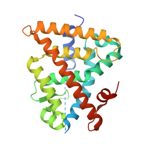
Structure-Guided Design of a Highly Potent Partial RXR Agonist with Superior Physicochemical Properties.

Lewandowski, M.Carmina, M.Knumann, L.Sai, M.Willems, S.Kasch, T.Pollinger, J.Knapp, S.Marschner, J.A.Chaikuad, A.Merk, D.

(2024) J Med Chem 67: 2152-2164

  • DOI: https://doi.org/10.1021/acs.jmedchem.3c02095
  • Primary Citation Related Structures: 
    8PP0

  • PubMed Abstract: 

    Retinoid X receptors (RXRs, NR2B1-3) hold therapeutic potential in oncology, neurodegeneration, and metabolic diseases, but traditional RXR agonists mimicking the natural ligand 9- cis retinoic acid exhibit poor physicochemical properties, pharmacokinetics, and safety profiles. Improved RXR ligands are needed to exploit RXR modulation as a promising therapeutic concept in various indications beyond its current role in second-line cancer treatment. Here, we report the co-crystal structure of RXR in complex with a novel pyrimidine-based ligand and the structure-informed optimization of this scaffold to highly potent and highly soluble RXR agonists. Focused structure-activity relationship elucidation and rigidization resulted in a substantially optimized partial RXR agonist with low nanomolar potency, no cytotoxic activity, and very favorable physicochemical properties highlighting this promising scaffold for the development of next-generation RXR targeting drugs.


  • Organizational Affiliation
    • Department of Pharmacy, Ludwig-Maximilians-Universität (LMU) München, 81377 Munich, Germany.

Macromolecule Content 

  • Total Structure Weight: 29.2 kDa 
  • Atom Count: 1,907 
  • Modeled Residue Count: 227 
  • Deposited Residue Count: 256 
  • Unique protein chains: 2

Macromolecules

Find similar proteins by:|  3D Structure
Entity ID: 1
MoleculeChains  Sequence LengthOrganismDetailsImage
Retinoic acid receptor RXR-alpha242Homo sapiensMutation(s): 0 
Gene Names: RXRANR2B1
UniProt & NIH Common Fund Data Resources
Find proteins for P19793 (Homo sapiens)
Explore P19793 
Go to UniProtKB:  P19793
PHAROS:  P19793
GTEx:  ENSG00000186350 
Entity Groups
Sequence Clusters30% Identity50% Identity70% Identity90% Identity95% Identity100% Identity
UniProt GroupP19793
Sequence Annotations
Expand
Reference Sequence
Find similar proteins by:  Sequence   |   3D Structure  
Entity ID: 2
MoleculeChains  Sequence LengthOrganismDetailsImage
Nuclear receptor coactivator 214Homo sapiensMutation(s): 0 
UniProt & NIH Common Fund Data Resources
Find proteins for Q15596 (Homo sapiens)
Explore Q15596 
Go to UniProtKB:  Q15596
PHAROS:  Q15596
GTEx:  ENSG00000140396 
Entity Groups
Sequence Clusters30% Identity50% Identity70% Identity90% Identity95% Identity100% Identity
UniProt GroupQ15596
Sequence Annotations
Expand
Reference Sequence

Small Molecules

Ligands 1 Unique
IDChains Name / Formula / InChI Key2D Diagram3D Interactions
7QJ
(Subject of Investigation/LOI)

Query on 7QJ


Download:Ideal Coordinates CCD File
C [auth A]3-[4-[2,3-dihydro-1H-inden-4-yl(methyl)amino]-6-(trifluoromethyl)pyrimidin-2-yl]oxypropanoic acid
C18 H18 F3 N3 O3
YMSJOQCCWIPKGF-UHFFFAOYSA-N
Binding Affinity Annotations 
IDSourceBinding Affinity
7QJ BindingDB:  8PP0 Kd: 95 (nM) from 1 assay(s)
EC50: min: 93, max: 360 (nM) from 3 assay(s)

Experimental Data & Validation

Experimental Data

  • Method: X-RAY DIFFRACTION
  • Resolution: 1.90 Å
  • R-Value Free:  0.223 (Depositor), 0.228 (DCC) 
  • R-Value Work:  0.180 (Depositor), 0.189 (DCC) 
  • R-Value Observed: 0.182 (Depositor) 
Space Group: P 43 21 2
Unit Cell:
Length ( Å )Angle ( ˚ )
a = 65.648α = 90
b = 65.648β = 90
c = 110.816γ = 90
Software Package:
Software NamePurpose
REFMACrefinement
Aimlessdata scaling
XDSdata reduction
PHASERphasing

Structure Validation

View Full Validation Report


Ligand Structure Quality Assessment 


Entry History 

& Funding Information

Deposition Data


Funding OrganizationLocationGrant Number
Not funded--

Revision History  (Full details and data files)

  • Version 1.0: 2024-02-07
    Type: Initial release
  • Version 1.1: 2024-02-21
    Changes: Database references