8P4T

The spike complex of the Lujo Virus


Experimental Data Snapshot

  • Method: ELECTRON MICROSCOPY
  • Resolution: 2.96 Å
  • Aggregation State: PARTICLE 
  • Reconstruction Method: SINGLE PARTICLE 

wwPDB Validation   3D Report Full Report


This is version 1.1 of the entry. See complete history


Literature

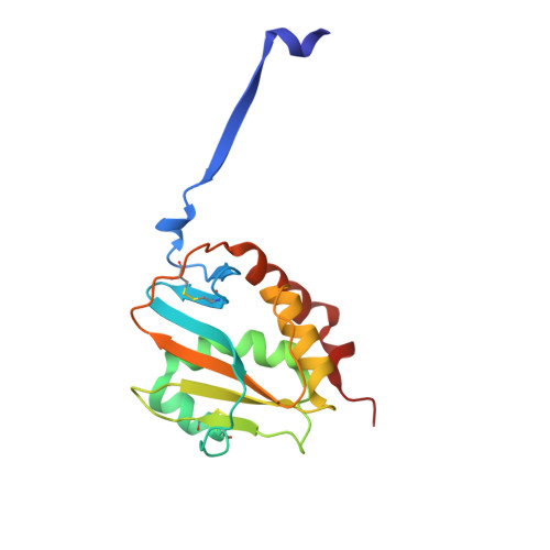
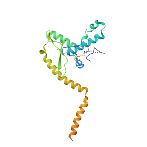
The structure of the Lujo virus spike complex.

Eilon-Ashkenazy, M.Cohen-Dvashi, H.Borni, S.Shaked, R.Calinsky, R.Levy, Y.Diskin, R.

(2024) Nat Commun 15: 7175-7175

  • DOI: https://doi.org/10.1038/s41467-024-51606-0
  • Primary Citation of Related Structures:  
    8P4T

  • PubMed Abstract: 

    Lujo virus (LUJV) is a human pathogen that was the cause of a deadly hemorrhagic fever outbreak in Africa. LUJV is a divergent member of the Arenaviridae with some similarities to both the "Old World" and "New World" serogroups, but it uses a cell-entry receptor, neuropilin-2 (NRP2), that is distinct from the receptors of OW and NW viruses. Though the receptor binding domain of LUJV has been characterized structurally, the overall organization of the trimeric spike complex and how NRP2 is recognized in this context were unknown. Here, we present the structure of the membrane-embedded LUJV spike complex determined by cryo-electron microscopy. Analysis of the structure suggested that a single NRP2 molecule is bound at the apex of the trimeric spike and that multiple subunits of the trimer contact the receptor. The binding of NRP2 involves an intriguing arginine-methionine interaction, which we analyzed using quantum mechanical modeling methods. We compare the LUJV spike structure with the only other available structure of a complete arenaviral spike, which is the Lassa virus. The similarities and differences between them shed light on Arenavirus evolution, inform vaccine design, and provide information that will be useful in combating future Arenavirus outbreaks.


  • Organizational Affiliation

    Department of Chemical and Structural Biology, Weizmann Institute of Science, Rehovot, Israel.


Macromolecules
Find similar proteins by:  (by identity cutoff)  |  3D Structure
Entity ID: 1
MoleculeChains Sequence LengthOrganismDetailsImage
GlycoproteinA,
D [auth B],
E [auth C]
163Mammarenavirus lujoenseMutation(s): 0 
UniProt
Find proteins for C5ILC1 (Mammarenavirus lujoense)
Explore C5ILC1 
Go to UniProtKB:  C5ILC1
Entity Groups  
Sequence Clusters30% Identity50% Identity70% Identity90% Identity95% Identity100% Identity
UniProt GroupC5ILC1
Glycosylation
Glycosylation Sites: 6
Sequence Annotations
Expand
  • Reference Sequence
Find similar proteins by:  (by identity cutoff)  |  3D Structure
Entity ID: 2
MoleculeChains Sequence LengthOrganismDetailsImage
GlycoproteinB [auth a],
H [auth b],
I [auth c]
247Mammarenavirus lujoenseMutation(s): 0 
UniProt
Find proteins for C5ILC1 (Mammarenavirus lujoense)
Explore C5ILC1 
Go to UniProtKB:  C5ILC1
Entity Groups  
Sequence Clusters30% Identity50% Identity70% Identity90% Identity95% Identity100% Identity
UniProt GroupC5ILC1
Glycosylation
Glycosylation Sites: 4
Sequence Annotations
Expand
  • Reference Sequence
Find similar proteins by:  (by identity cutoff)  |  3D Structure
Entity ID: 3
MoleculeChains Sequence LengthOrganismDetailsImage
GlycoproteinC [auth SA],
F [auth SB],
G [auth SC]
58Mammarenavirus lujoenseMutation(s): 0 
UniProt
Find proteins for C5ILC1 (Mammarenavirus lujoense)
Explore C5ILC1 
Go to UniProtKB:  C5ILC1
Entity Groups  
Sequence Clusters30% Identity50% Identity70% Identity90% Identity95% Identity100% Identity
UniProt GroupC5ILC1
Sequence Annotations
Expand
  • Reference Sequence
Oligosaccharides

Help

Entity ID: 4
MoleculeChains Length2D Diagram Glycosylation3D Interactions
alpha-D-mannopyranose-(1-3)-beta-D-mannopyranose-(1-4)-2-acetamido-2-deoxy-beta-D-glucopyranose-(1-4)-[alpha-L-fucopyranose-(1-6)]2-acetamido-2-deoxy-beta-D-glucopyranoseJ [auth D],
M [auth G],
O [auth I]
5N-Glycosylation
Glycosylation Resources
GlyTouCan:  G42466VF
GlyCosmos:  G42466VF
GlyGen:  G42466VF
Entity ID: 5
MoleculeChains Length2D Diagram Glycosylation3D Interactions
alpha-D-mannopyranose-(1-2)-alpha-D-mannopyranose-(1-3)-[alpha-D-mannopyranose-(1-6)-alpha-D-mannopyranose-(1-6)]beta-D-mannopyranose-(1-4)-2-acetamido-2-deoxy-beta-D-glucopyranose-(1-4)-2-acetamido-2-deoxy-beta-D-glucopyranoseK [auth E],
N [auth H],
P [auth J]
7N-Glycosylation
Glycosylation Resources
GlyTouCan:  G23799GS
GlyCosmos:  G23799GS
GlyGen:  G23799GS
Entity ID: 6
MoleculeChains Length2D Diagram Glycosylation3D Interactions
2-acetamido-2-deoxy-beta-D-glucopyranose-(1-4)-2-acetamido-2-deoxy-beta-D-glucopyranoseL [auth F],
Q [auth K],
R [auth L]
2N-Glycosylation
Glycosylation Resources
GlyTouCan:  G42666HT
GlyCosmos:  G42666HT
GlyGen:  G42666HT
Small Molecules
Ligands 2 Unique
IDChains Name / Formula / InChI Key2D Diagram3D Interactions
NAG
Query on NAG

Download Ideal Coordinates CCD File 
AA [auth B]
BA [auth B]
CA [auth B]
DA [auth B]
EA [auth C]
AA [auth B],
BA [auth B],
CA [auth B],
DA [auth B],
EA [auth C],
FA [auth C],
GA [auth C],
HA [auth C],
IA [auth b],
JA [auth b],
KA [auth b],
LA [auth c],
MA [auth c],
NA [auth c],
S [auth A],
T [auth A],
U [auth A],
V [auth A],
X [auth a],
Y [auth a],
Z [auth a]
2-acetamido-2-deoxy-beta-D-glucopyranose
C8 H15 N O6
OVRNDRQMDRJTHS-FMDGEEDCSA-N
NA
Query on NA

Download Ideal Coordinates CCD File 
W [auth A]SODIUM ION
Na
FKNQFGJONOIPTF-UHFFFAOYSA-N
Experimental Data & Validation

Experimental Data

  • Method: ELECTRON MICROSCOPY
  • Resolution: 2.96 Å
  • Aggregation State: PARTICLE 
  • Reconstruction Method: SINGLE PARTICLE 
EM Software:
TaskSoftware PackageVersion
MODEL REFINEMENTPHENIX1.21.1_5286

Structure Validation

View Full Validation Report


Entry History & Funding Information

Deposition Data


Funding OrganizationLocationGrant Number
Israel Science FoundationIsrael209/20

Revision History  (Full details and data files)

  • Version 1.0: 2024-06-12
    Type: Initial release
  • Version 1.1: 2024-09-04
    Changes: Data collection, Database references