8OWY

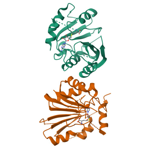
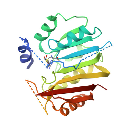
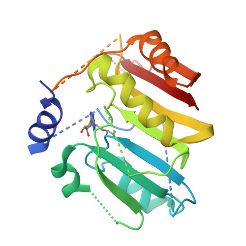
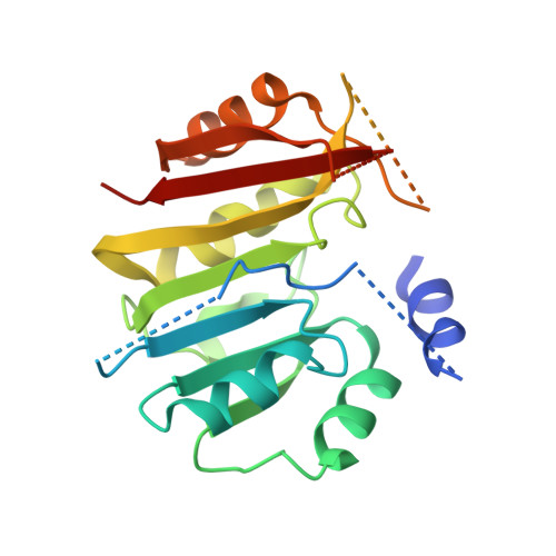
Crystal structure of METTL6 mutant 40-269 bound to SAH


Experimental Data Snapshot

  • Method: X-RAY DIFFRACTION
  • Resolution: 3.20 Å
  • R-Value Free: 0.300 
  • R-Value Work: 0.260 
  • R-Value Observed: 0.262 

Starting Model: experimental
View more details

wwPDB Validation   3D Report Full Report


Ligand Structure Quality Assessment 


This is version 1.4 of the entry. See complete history


Literature

Structural basis of tRNA recognition by the m 3 C RNA methyltransferase METTL6 in complex with SerRS seryl-tRNA synthetase.

Throll, P.G Dolce, L.Rico-Lastres, P.Arnold, K.Tengo, L.Basu, S.Kaiser, S.Schneider, R.Kowalinski, E.

(2024) Nat Struct Mol Biol 31: 1614-1624

  • DOI: https://doi.org/10.1038/s41594-024-01341-3
  • Primary Citation of Related Structures:  
    8OWX, 8OWY, 8P7B, 8P7C, 8P7D

  • PubMed Abstract: 

    Methylation of cytosine 32 in the anticodon loop of tRNAs to 3-methylcytosine (m 3 C) is crucial for cellular translation fidelity. Misregulation of the RNA methyltransferases setting this modification can cause aggressive cancers and metabolic disturbances. Here, we report the cryo-electron microscopy structure of the human m 3 C tRNA methyltransferase METTL6 in complex with seryl-tRNA synthetase (SerRS) and their common substrate tRNA Ser . Through the complex structure, we identify the tRNA-binding domain of METTL6. We show that SerRS acts as the tRNA Ser substrate selection factor for METTL6. We demonstrate that SerRS augments the methylation activity of METTL6 and that direct contacts between METTL6 and SerRS are necessary for efficient tRNA Ser methylation. Finally, on the basis of the structure of METTL6 in complex with SerRS and tRNA Ser , we postulate a universal tRNA-binding mode for m 3 C RNA methyltransferases, including METTL2 and METTL8, suggesting that these mammalian paralogs use similar ways to engage their respective tRNA substrates and cofactors.


  • Organizational Affiliation

    European Molecular Biology Laboratory, Grenoble, France.


Macromolecules
Find similar proteins by:  (by identity cutoff)  |  3D Structure
Entity ID: 1
MoleculeChains Sequence LengthOrganismDetailsImage
tRNA N(3)-methylcytidine methyltransferase METTL6
A, B
235Homo sapiensMutation(s): 0 
Gene Names: METTL6
EC: 2.1.1
UniProt & NIH Common Fund Data Resources
Find proteins for Q8TCB7 (Homo sapiens)
Explore Q8TCB7 
Go to UniProtKB:  Q8TCB7
PHAROS:  Q8TCB7
GTEx:  ENSG00000206562 
Entity Groups  
Sequence Clusters30% Identity50% Identity70% Identity90% Identity95% Identity100% Identity
UniProt GroupQ8TCB7
Sequence Annotations
Expand
  • Reference Sequence
Experimental Data & Validation

Experimental Data

  • Method: X-RAY DIFFRACTION
  • Resolution: 3.20 Å
  • R-Value Free: 0.300 
  • R-Value Work: 0.260 
  • R-Value Observed: 0.262 
  • Space Group: C 1 2 1
Unit Cell:
Length ( Å )Angle ( ˚ )
a = 81.971α = 90
b = 138.129β = 115.26
c = 39.66γ = 90
Software Package:
Software NamePurpose
PHENIXrefinement
Cootmodel building
XDSdata reduction
XSCALEdata scaling
PHENIXphasing

Structure Validation

View Full Validation Report


Ligand Structure Quality Assessment 


Entry History & Funding Information

Deposition Data


Funding OrganizationLocationGrant Number
Other government--

Revision History  (Full details and data files)

  • Version 1.0: 2024-05-08
    Type: Initial release
  • Version 1.1: 2024-05-22
    Changes: Refinement description
  • Version 1.2: 2024-07-03
    Changes: Database references
  • Version 1.3: 2024-07-10
    Changes: Database references
  • Version 1.4: 2024-10-30
    Changes: Data collection, Database references, Structure summary