8OW6 | pdb_00008ow6

PERIDININ-CHLOROPHYLL-PROTEIN OF HETEROCAPSA PYGMAEA, 100K


Experimental Data Snapshot

  • Method: X-RAY DIFFRACTION
  • Resolution: 1.20 Å
  • R-Value Free: 
    0.171 (Depositor), 0.165 (DCC) 
  • R-Value Work: 
    0.149 (Depositor) 
  • R-Value Observed: 
    0.163 (Depositor) 

Starting Model: experimental
View more details

wwPDB Validation 3D Report Full Report

Validation slider image for 8OW6

Ligand Structure Quality Assessment 


This is version 1.2 of the entry. See complete history

Literature

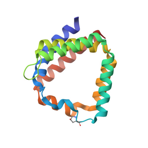
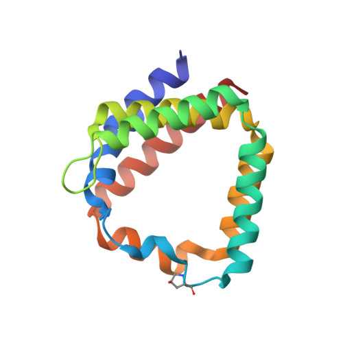
Structural and spectroscopic characterization of the peridinin-chlorophyll a-protein (PCP) complex from Heterocapsa pygmaea (HPPCP).

Schulte, T.Magdaong, N.C.M.Di Valentin, M.Agostini, A.Tait, C.E.Niedzwiedzki, D.M.Carbonera, D.Hofmann, E.

(2024) Biochim Biophys Acta Bioenerg 1866: 149510-149510

  • DOI: https://doi.org/10.1016/j.bbabio.2024.149510
  • Primary Citation Related Structures: 
    8OV5, 8OW6

  • PubMed Abstract: 

    Light harvesting proteins are optimized to efficiently collect and transfer light energy for photosynthesis. In eukaryotic dinoflagellates these complexes utilize chlorophylls and a special carotenoid, peridinin, and arrange them for efficient excitation energy transfer. At the same time, the carotenoids protect the system by quenching harmful chlorophyll triplet states. Here we use advanced spectroscopic techniques and X-ray structure analysis to investigate excitation energy transfer processes in the major soluble antenna, the peridinin chlorophyll a protein (PCP) from the free living dinoflagellate Heterocapsa pygmaea. We determined the 3D-structure of this complex at high resolution (1.2 Å). For better comparison, we improved the reference structure of this protein from Amphidinium carterae to a resolution of 1.15 Å. We then used fs and ns time-resolved absorption spectroscopy to study the mechanisms of light harvesting, but also of the photoprotective quenching of the chlorophyll triplet state. The photoprotection site was further characterized by Electron Spin Echo Envelope Modulation (ESEEM) spectroscopy to yield information on water molecules involved in triplet-triplet energy transfer. Similar to other PCP complexes, excitation energy transfer from peridinin to chlorophyll is found to be very efficient, with transfer times in the range of 1.6-2.1 ps. One of the four carotenoids, the peridinin 614, is well positioned to quench the chlorophyll triplet state with high efficiency and transfer times in the range of tens of picoseconds. Our structural and dynamic data further support, that the intrinsic water molecule coordinating the chlorophyll Mg ion plays an essential role in photoprotection.


  • Organizational Affiliation
    • Protein Crystallography, Faculty of Biology and Biotechnology, Ruhr University Bochum, 44801 Bochum, Germany; Department of Biochemistry and Biophysics, National Bioinformatics Infrastructure Sweden, Science for Life Laboratory, Stockholm University, 17121 Solna, Sweden.

Macromolecule Content 

  • Total Structure Weight: 80.37 kDa 
  • Atom Count: 6,601 
  • Modeled Residue Count: 596 
  • Deposited Residue Count: 596 
  • Unique protein chains: 2

Macromolecules

Find similar proteins by:|  3D Structure
Entity ID: 1
MoleculeChains  Sequence LengthOrganismDetailsImage
Peridinin-chl a protein
A, C
149Heterocapsa pygmaeaMutation(s): 0 
UniProt
Find proteins for Q9FEY4 (Heterocapsa pygmaea)
Explore Q9FEY4 
Go to UniProtKB:  Q9FEY4
Entity Groups
Sequence Clusters30% Identity50% Identity70% Identity90% Identity95% Identity100% Identity
UniProt GroupQ9FEY4
Sequence Annotations
Expand
Reference Sequence
Find similar proteins by:|  3D Structure
Entity ID: 2
MoleculeChains  Sequence LengthOrganismDetailsImage
Peridinin-chl a protein
B, D
149Heterocapsa pygmaeaMutation(s): 0 
UniProt
Find proteins for Q9FEY4 (Heterocapsa pygmaea)
Explore Q9FEY4 
Go to UniProtKB:  Q9FEY4
Entity Groups
Sequence Clusters30% Identity50% Identity70% Identity90% Identity95% Identity100% Identity
UniProt GroupQ9FEY4
Sequence Annotations
Expand
Reference Sequence

Small Molecules

Ligands 6 Unique
IDChains Name / Formula / InChI Key2D Diagram3D Interactions
W4O
(Subject of Investigation/LOI)

Query on W4O


Download:Ideal Coordinates CCD File
FA [auth D],
J [auth A],
R [auth B],
Z [auth C]
[(2~{S})-3-[(2~{R},3~{R},4~{S},5~{R},6~{R})-6-[[(2~{S},3~{R},4~{S},5~{R},6~{R})-6-(hydroxymethyl)-3,4,5-tris(oxidanyl)oxan-2-yl]oxymethyl]-3,4,5-tris(oxidanyl)oxan-2-yl]oxy-2-[(6~{Z},9~{Z},12~{Z},15~{Z})-octadeca-6,9,12,15-tetraenoyl]oxy-propyl] (6~{Z},9~{Z},12~{Z},15~{Z})-octadeca-6,9,12,15-tetraenoate
C51 H80 O15
VXOQBABFAYTJKD-LAAVQGTGSA-N
CLA
(Subject of Investigation/LOI)

Query on CLA


Download:Ideal Coordinates CCD File
AA [auth D],
E [auth A],
M [auth B],
U [auth C]
CHLOROPHYLL A
C55 H72 Mg N4 O5
ATNHDLDRLWWWCB-AENOIHSZSA-M
PID
(Subject of Investigation/LOI)

Query on PID


Download:Ideal Coordinates CCD File
BA [auth D]
CA [auth D]
DA [auth D]
EA [auth D]
F [auth A]
BA [auth D],
CA [auth D],
DA [auth D],
EA [auth D],
F [auth A],
G [auth A],
H [auth A],
I [auth A],
N [auth B],
O [auth B],
P [auth B],
Q [auth B],
V [auth C],
W [auth C],
X [auth C],
Y [auth C]
PERIDININ
C39 H50 O7
UYRDHEJRPVSJFM-FROCQLDGSA-N
PO4

Query on PO4


Download:Ideal Coordinates CCD File
L [auth A]PHOSPHATE ION
O4 P
NBIIXXVUZAFLBC-UHFFFAOYSA-K
K

Query on K


Download:Ideal Coordinates CCD File
K [auth A],
S [auth B]
POTASSIUM ION
K
NPYPAHLBTDXSSS-UHFFFAOYSA-N
CL

Query on CL


Download:Ideal Coordinates CCD File
T [auth B]CHLORIDE ION
Cl
VEXZGXHMUGYJMC-UHFFFAOYSA-M
Modified Residues  1 Unique
IDChains TypeFormula2D DiagramParent
HYP
Query on HYP
A, C
L-PEPTIDE LINKINGC5 H9 N O3PRO

Experimental Data & Validation

Experimental Data

  • Method: X-RAY DIFFRACTION
  • Resolution: 1.20 Å
  • R-Value Free:  0.171 (Depositor), 0.165 (DCC) 
  • R-Value Work:  0.149 (Depositor) 
  • R-Value Observed: 0.163 (Depositor) 
Space Group: P 3 1 2
Unit Cell:
Length ( Å )Angle ( ˚ )
a = 90.61α = 90
b = 90.61β = 90
c = 155.61γ = 120
Software Package:
Software NamePurpose
PHENIXrefinement
XDSdata reduction
XSCALEdata scaling
PHASERphasing

Structure Validation

View Full Validation Report


Ligand Structure Quality Assessment 


Entry History 

& Funding Information

Deposition Data


Funding OrganizationLocationGrant Number
German Research Foundation (DFG)GermanySFB480

Revision History  (Full details and data files)

  • Version 1.0: 2024-05-15
    Type: Initial release
  • Version 1.1: 2024-10-09
    Changes: Database references, Structure summary
  • Version 1.2: 2024-10-16
    Changes: Database references