8OFV | pdb_00008ofv

Human adenovirus type 53 fiber-knob protein complexed with sialic acid


Experimental Data Snapshot

  • Method: X-RAY DIFFRACTION
  • Resolution: 1.77 Å
  • R-Value Free: 
    0.228 (Depositor), 0.198 (DCC) 
  • R-Value Work: 
    0.208 (Depositor) 
  • R-Value Observed: 
    0.209 (Depositor) 

wwPDB Validation 3D Report Full Report

Validation slider image for 8OFV

Ligand Structure Quality Assessment 


This is version 1.3 of the entry. See complete history

Literature

Broad sialic acid usage amongst species D human adenovirus.

Mundy, R.M.Baker, A.T.Bates, E.A.Cunliffe, T.G.Teijeira-Crespo, A.Moses, E.Rizkallah, P.J.Parker, A.L.

(2023) Npj Viruses 1: 1-1

  • DOI: https://doi.org/10.1038/s44298-023-00001-5
  • Primary Citation Related Structures: 
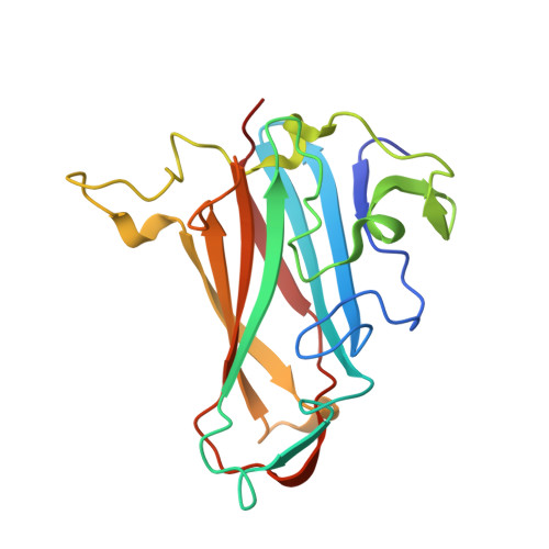
    8OFP, 8OFQ, 8OFR, 8OFS, 8OFT, 8OFU, 8OFV

  • PubMed Abstract: 

    Human adenoviruses (HAdV) are widespread pathogens causing usually mild infections. The Species D (HAdV-D) cause gastrointestinal tract infections and epidemic keratoconjunctivitis (EKC). Despite being significant pathogens, knowledge around HAdV-D mechanism of cell infection is lacking. Sialic acid (SA) usage has been proposed as a cell infection mechanism for EKC causing HAdV-D. Here we highlight an important role for SA engagement by many HAdV-D. We provide apo state crystal structures of 7 previously undetermined HAdV-D fiber-knob proteins, and structures of HAdV-D25, D29, D30 and D53 fiber-knob proteins in complex with SA. Biologically, we demonstrate that removal of cell surface SA reduced infectivity of HAdV-C5 vectors pseudotyped with HAdV-D fiber-knob proteins, whilst engagement of the classical HAdV receptor CAR was variable. Our data indicates variable usage of SA and CAR across HAdV-D. Better defining these interactions will enable improved development of antivirals and engineering of the viruses into refined therapeutic vectors.


  • Organizational Affiliation
    • Division of Cancer & Genetics, Cardiff University School of Medicine, Cardiff, CF14 4XN UK.

Macromolecule Content 

  • Total Structure Weight: 260.9 kDa 
  • Atom Count: 19,866 
  • Modeled Residue Count: 2,214 
  • Deposited Residue Count: 2,268 
  • Unique protein chains: 1

Macromolecules

Find similar proteins by:|  3D Structure
Entity ID: 1
MoleculeChains  Sequence LengthOrganismDetailsImage
Fiber protein
A, B, C, D, E
A, B, C, D, E, F, G, H, I, J, K, L
189Human adenovirus 53Mutation(s): 0 
Gene Names: L5
UniProt
Find proteins for E5RWD1 (Human adenovirus 53)
Explore E5RWD1 
Go to UniProtKB:  E5RWD1
Entity Groups
Sequence Clusters30% Identity50% Identity70% Identity90% Identity95% Identity100% Identity
UniProt GroupE5RWD1
Sequence Annotations
Expand
Reference Sequence

Small Molecules

Ligands 5 Unique
IDChains Name / Formula / InChI Key2D Diagram3D Interactions
SIA

Query on SIA


Download:Ideal Coordinates CCD File
BB [auth L]N-acetyl-alpha-neuraminic acid
C11 H19 N O9
SQVRNKJHWKZAKO-YRMXFSIDSA-N
SLB
(Subject of Investigation/LOI)

Query on SLB


Download:Ideal Coordinates CCD File
HA [auth F]N-acetyl-beta-neuraminic acid
C11 H19 N O9
SQVRNKJHWKZAKO-PFQGKNLYSA-N
P4G

Query on P4G


Download:Ideal Coordinates CCD File
AB [auth L]
GA [auth F]
NA [auth G]
OA [auth G]
V [auth B]
AB [auth L],
GA [auth F],
NA [auth G],
OA [auth G],
V [auth B],
VA [auth I],
W [auth B],
XA [auth K],
Y [auth C],
Z [auth C],
ZA [auth L]
1-ETHOXY-2-(2-ETHOXYETHOXY)ETHANE
C8 H18 O3
RRQYJINTUHWNHW-UHFFFAOYSA-N
PEG

Query on PEG


Download:Ideal Coordinates CCD File
BA [auth E]
CA [auth E]
DA [auth E]
FA [auth F]
LA [auth G]
BA [auth E],
CA [auth E],
DA [auth E],
FA [auth F],
LA [auth G],
M [auth A],
MA [auth G],
Q [auth B],
QA [auth H],
R [auth B],
RA [auth H],
S [auth B],
T [auth B],
TA [auth I],
U [auth B],
UA [auth I],
WA [auth K],
X [auth C]
DI(HYDROXYETHYL)ETHER
C4 H10 O3
MTHSVFCYNBDYFN-UHFFFAOYSA-N
EDO

Query on EDO


Download:Ideal Coordinates CCD File
AA [auth E]
EA [auth F]
IA [auth G]
JA [auth G]
KA [auth G]
AA [auth E],
EA [auth F],
IA [auth G],
JA [auth G],
KA [auth G],
N [auth B],
O [auth B],
P [auth B],
PA [auth H],
SA [auth I],
YA [auth L]
1,2-ETHANEDIOL
C2 H6 O2
LYCAIKOWRPUZTN-UHFFFAOYSA-N

Experimental Data & Validation

Experimental Data

  • Method: X-RAY DIFFRACTION
  • Resolution: 1.77 Å
  • R-Value Free:  0.228 (Depositor), 0.198 (DCC) 
  • R-Value Work:  0.208 (Depositor) 
  • R-Value Observed: 0.209 (Depositor) 
Space Group: P 1 2 1
Unit Cell:
Length ( Å )Angle ( ˚ )
a = 93.555α = 90
b = 60.406β = 99.88
c = 243.686γ = 90
Software Package:
Software NamePurpose
REFMACrefinement
DIALSdata scaling
xia2data reduction
PHASERphasing

Structure Validation

View Full Validation Report


Ligand Structure Quality Assessment 


Entry History 

& Funding Information

Deposition Data


Funding OrganizationLocationGrant Number
Medical Research Council (MRC, United Kingdom)United KingdomMR/N0137941/1
Wellcome TrustUnited Kingdom517732

Revision History  (Full details and data files)

  • Version 1.0: 2023-09-20
    Type: Initial release
  • Version 1.1: 2023-10-11
    Changes: Database references
  • Version 1.2: 2024-05-08
    Changes: Database references
  • Version 1.3: 2024-11-06
    Changes: Structure summary