8OE2 | pdb_00008oe2

Structure of hyperstable haloalkane dehalogenase variant DhaA223


Experimental Data Snapshot

  • Method: X-RAY DIFFRACTION
  • Resolution: 1.51 Å
  • R-Value Free: 
    0.195 (Depositor), 0.194 (DCC) 
  • R-Value Work: 
    0.168 (Depositor), 0.169 (DCC) 
  • R-Value Observed: 
    0.170 (Depositor) 

Starting Model: experimental
View more details

wwPDB Validation 3D Report Full Report

Validation slider image for 8OE2

Ligand Structure Quality Assessment 


This is version 1.0 of the entry. See complete history

Literature

Advancing Enzyme's Stability and Catalytic Efficiency through Synergy of Force-Field Calculations, Evolutionary Analysis, and Machine Learning.

Kunka, A.Marques, S.M.Havlasek, M.Vasina, M.Velatova, N.Cengelova, L.Kovar, D.Damborsky, J.Marek, M.Bednar, D.Prokop, Z.

(2023) ACS Catal 13: 12506-12518

  • DOI: https://doi.org/10.1021/acscatal.3c02575
  • Primary Citation Related Structures: 
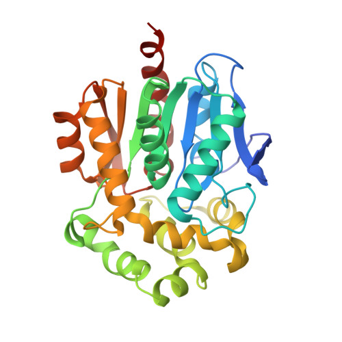
    8OE2, 8OE6

  • PubMed Abstract: 

    Thermostability is an essential requirement for the use of enzymes in the bioindustry. Here, we compare different protein stabilization strategies using a challenging target, a stable haloalkane dehalogenase DhaA115. We observe better performance of automated stabilization platforms FireProt and PROSS in designing multiple-point mutations over the introduction of disulfide bonds and strengthening the intra- and the inter-domain contacts by in silico saturation mutagenesis. We reveal that the performance of automated stabilization platforms was still compromised due to the introduction of some destabilizing mutations. Notably, we show that their prediction accuracy can be improved by applying manual curation or machine learning for the removal of potentially destabilizing mutations, yielding highly stable haloalkane dehalogenases with enhanced catalytic properties. A comparison of crystallographic structures revealed that current stabilization rounds were not accompanied by large backbone re-arrangements previously observed during the engineering stability of DhaA115. Stabilization was achieved by improving local contacts including protein-water interactions. Our study provides guidance for further improvement of automated structure-based computational tools for protein stabilization.


  • Organizational Affiliation
    • Loschmidt Laboratories, Department of Experimental Biology and RECETOX, Faculty of Science, Masaryk University, Brno 601 77, Czech Republic.

Macromolecule Content 

  • Total Structure Weight: 210.55 kDa 
  • Atom Count: 16,472 
  • Modeled Residue Count: 1,748 
  • Deposited Residue Count: 1,794 
  • Unique protein chains: 1

Macromolecules

Find similar proteins by:|  3D Structure
Entity ID: 1
MoleculeChains  Sequence LengthOrganismDetailsImage
Haloalkane dehalogenase
A, B, C, D, E
A, B, C, D, E, F
299synthetic constructMutation(s): 0 
Gene Names: dhaA
EC: 3.8.1.5
UniProt
Find proteins for P0A3G4 (Pseudomonas pavonaceae)
Explore P0A3G4 
Go to UniProtKB:  P0A3G4
Entity Groups
Sequence Clusters30% Identity50% Identity70% Identity90% Identity95% Identity100% Identity
UniProt GroupP0A3G4
Sequence Annotations
Expand
Reference Sequence

Small Molecules

Ligands 5 Unique
IDChains Name / Formula / InChI Key2D Diagram3D Interactions
PG4
(Subject of Investigation/LOI)

Query on PG4


Download:Ideal Coordinates CCD File
FA [auth E],
I [auth A],
Z [auth D]
TETRAETHYLENE GLYCOL
C8 H18 O5
UWHCKJMYHZGTIT-UHFFFAOYSA-N
TRS
(Subject of Investigation/LOI)

Query on TRS


Download:Ideal Coordinates CCD File
L [auth A]2-AMINO-2-HYDROXYMETHYL-PROPANE-1,3-DIOL
C4 H12 N O3
LENZDBCJOHFCAS-UHFFFAOYSA-O
SO4
(Subject of Investigation/LOI)

Query on SO4


Download:Ideal Coordinates CCD File
CA [auth E]
H [auth A]
P [auth B]
T [auth C]
U [auth C]
CA [auth E],
H [auth A],
P [auth B],
T [auth C],
U [auth C],
X [auth D],
Y [auth D]
SULFATE ION
O4 S
QAOWNCQODCNURD-UHFFFAOYSA-L
GOL
(Subject of Investigation/LOI)

Query on GOL


Download:Ideal Coordinates CCD File
AA [auth D]
DA [auth E]
EA [auth E]
GA [auth E]
IA [auth F]
AA [auth D],
DA [auth E],
EA [auth E],
GA [auth E],
IA [auth F],
J [auth A],
K [auth A],
M [auth A],
N [auth A],
Q [auth B],
R [auth B],
V [auth C]
GLYCEROL
C3 H8 O3
PEDCQBHIVMGVHV-UHFFFAOYSA-N
CL
(Subject of Investigation/LOI)

Query on CL


Download:Ideal Coordinates CCD File
BA [auth E]
G [auth A]
HA [auth F]
O [auth B]
S [auth C]
BA [auth E],
G [auth A],
HA [auth F],
O [auth B],
S [auth C],
W [auth D]
CHLORIDE ION
Cl
VEXZGXHMUGYJMC-UHFFFAOYSA-M

Experimental Data & Validation

Experimental Data

  • Method: X-RAY DIFFRACTION
  • Resolution: 1.51 Å
  • R-Value Free:  0.195 (Depositor), 0.194 (DCC) 
  • R-Value Work:  0.168 (Depositor), 0.169 (DCC) 
  • R-Value Observed: 0.170 (Depositor) 
Space Group: P 1 21 1
Unit Cell:
Length ( Å )Angle ( ˚ )
a = 67.35α = 90
b = 143.144β = 108.38
c = 106.676γ = 90
Software Package:
Software NamePurpose
PHENIXrefinement
Aimlessdata scaling
XDSdata reduction
PHASERphasing

Structure Validation

View Full Validation Report


Ligand Structure Quality Assessment 


Entry History 

& Funding Information

Deposition Data

  • Released Date: 2024-01-17 
  • Deposition Author(s): Marek, M.

Funding OrganizationLocationGrant Number
Czech Science FoundationCzech RepublicGA22-09853S

Revision History  (Full details and data files)

  • Version 1.0: 2024-01-17
    Type: Initial release