Identification of enzymatic functions of osmo-regulated periplasmic glucan biosynthesis proteins from Escherichia coli reveals a novel glycoside hydrolase family.
Motouchi, S., Kobayashi, K., Nakai, H., Nakajima, M.(2023) Commun Biol 6: 961-961
- PubMed: 37735577 Search on PubMedSearch on PubMed Central
- DOI: https://doi.org/10.1038/s42003-023-05336-6
- Primary Citation Related Structures:
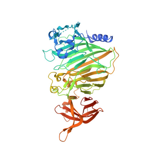
8IOX, 8IP1, 8IP2 - PubMed Abstract:
Most Gram-negative bacteria synthesize osmo-regulated periplasmic glucans (OPG) in the periplasm or extracellular space. Pathogenicity of many pathogens is lost by knocking out opgG, an OPG-related gene indispensable for OPG synthesis. However, the biochemical functions of OpgG and OpgD, a paralog of OpgG, have not been elucidated. In this study, structural and functional analyses of OpgG and OpgD from Escherichia coli revealed that these proteins are β-1,2-glucanases with remarkably different activity from each other, establishing a new glycoside hydrolase family, GH186. Furthermore, a reaction mechanism with an unprecedentedly long proton transfer pathway among glycoside hydrolase families is proposed for OpgD. The conformation of the region that forms the reaction pathway differs noticeably between OpgG and OpgD, which explains the observed low activity of OpgG. The findings enhance our understanding of OPG biosynthesis and provide insights into functional diversity for this novel enzyme family.
- Department of Applied Biological Science, Faculty of Science and Technology, Tokyo University of Science, 2641 Yamazaki, Noda Chiba, 278-8510, Japan.
Organizational Affiliation: