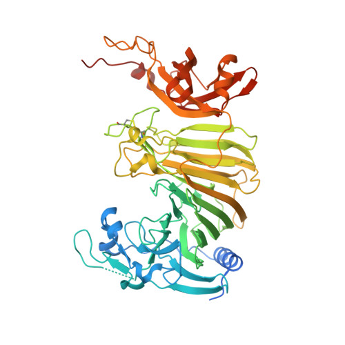
8IP1

Escherichia coli OpgD mutant-D388N with beta-1,2-glucan


Experimental Data Snapshot

  • Method: X-RAY DIFFRACTION
  • Resolution: 2.06 Å
  • R-Value Free: 0.224 
  • R-Value Work: 0.175 

wwPDB Validation   3D Report Full Report


This is version 1.2 of the entry. See complete history


Literature

Identification of enzymatic functions of osmo-regulated periplasmic glucan biosynthesis proteins from Escherichia coli reveals a novel glycoside hydrolase family.

Motouchi, S.Kobayashi, K.Nakai, H.Nakajima, M.

(2023) Commun Biol 6: 961-961

  • DOI: https://doi.org/10.1038/s42003-023-05336-6
  • Primary Citation of Related Structures:  
    8IOX, 8IP1, 8IP2

  • PubMed Abstract: 

    Most Gram-negative bacteria synthesize osmo-regulated periplasmic glucans (OPG) in the periplasm or extracellular space. Pathogenicity of many pathogens is lost by knocking out opgG, an OPG-related gene indispensable for OPG synthesis. However, the biochemical functions of OpgG and OpgD, a paralog of OpgG, have not been elucidated. In this study, structural and functional analyses of OpgG and OpgD from Escherichia coli revealed that these proteins are β-1,2-glucanases with remarkably different activity from each other, establishing a new glycoside hydrolase family, GH186. Furthermore, a reaction mechanism with an unprecedentedly long proton transfer pathway among glycoside hydrolase families is proposed for OpgD. The conformation of the region that forms the reaction pathway differs noticeably between OpgG and OpgD, which explains the observed low activity of OpgG. The findings enhance our understanding of OPG biosynthesis and provide insights into functional diversity for this novel enzyme family.


  • Organizational Affiliation

    Department of Applied Biological Science, Faculty of Science and Technology, Tokyo University of Science, 2641 Yamazaki, Noda Chiba, 278-8510, Japan.


Macromolecules
Find similar proteins by:  (by identity cutoff)  |  3D Structure
Entity ID: 1
MoleculeChains Sequence LengthOrganismDetailsImage
Glucans biosynthesis protein D
A, B
559Escherichia coliMutation(s): 1 
Gene Names: opgD
UniProt
Find proteins for P40120 (Escherichia coli (strain K12))
Explore P40120 
Go to UniProtKB:  P40120
Entity Groups  
Sequence Clusters30% Identity50% Identity70% Identity90% Identity95% Identity100% Identity
UniProt GroupP40120
Sequence Annotations
Expand
  • Reference Sequence
Oligosaccharides

Help

Entity ID: 2
MoleculeChains Length2D Diagram Glycosylation3D Interactions
beta-D-glucopyranose-(1-2)-beta-D-glucopyranose-(1-2)-beta-D-glucopyranose-(1-2)-beta-D-glucopyranose-(1-2)-beta-D-glucopyranose-(1-2)-beta-D-glucopyranose-(1-2)-beta-D-glucopyranose-(1-2)-beta-D-glucopyranose-(1-2)-beta-D-glucopyranose-(1-2)-beta-D-glucopyranose-(1-2)-beta-D-glucopyranose
C
11N/A
Entity ID: 3
MoleculeChains Length2D Diagram Glycosylation3D Interactions
beta-D-glucopyranose-(1-2)-beta-D-glucopyranose-(1-2)-beta-D-glucopyranose-(1-2)-beta-D-glucopyranose-(1-2)-beta-D-glucopyranose-(1-2)-beta-D-glucopyranose-(1-2)-beta-D-glucopyranose-(1-2)-beta-D-glucopyranose-(1-2)-beta-D-glucopyranose-(1-2)-beta-D-glucopyranose-(1-2)-beta-D-glucopyranose-(1-2)-beta-D-glucopyranose-(1-2)-beta-D-glucopyranoseD [auth E]13N/A
Experimental Data & Validation

Experimental Data

  • Method: X-RAY DIFFRACTION
  • Resolution: 2.06 Å
  • R-Value Free: 0.224 
  • R-Value Work: 0.175 
  • Space Group: P 1 21 1
Unit Cell:
Length ( Å )Angle ( ˚ )
a = 58.092α = 90
b = 87.099β = 101.132
c = 110.853γ = 90
Software Package:
Software NamePurpose
REFMACrefinement
XDSdata reduction
Aimlessdata scaling
MOLREPphasing

Structure Validation

View Full Validation Report


Entry History & Funding Information

Deposition Data


Funding OrganizationLocationGrant Number
Japan Society for the Promotion of Science (JSPS)Japan--

Revision History  (Full details and data files)

  • Version 1.0: 2024-03-13
    Type: Initial release
  • Version 1.1: 2024-07-10
    Changes: Database references
  • Version 1.2: 2024-07-17
    Changes: Database references