8HZV | pdb_00008hzv

The crystal structure of a Radical SAM Enzyme DesII


Experimental Data Snapshot

  • Method: X-RAY DIFFRACTION
  • Resolution: 2.33 Å
  • R-Value Free: 
    0.235 (Depositor), 0.237 (DCC) 
  • R-Value Work: 
    0.204 (Depositor), 0.206 (DCC) 
  • R-Value Observed: 
    0.206 (Depositor) 

wwPDB Validation 3D Report Full Report

Validation slider image for 8HZV

Ligand Structure Quality Assessment 


This is version 1.2 of the entry. See complete history

Literature

Mechanistic Insights from the Crystal Structure and Computational Analysis of the Radical SAM Deaminase DesII.

Hou, X.Feng, J.Franklin, J.L.Russo, R.Guo, Z.Zhou, J.Gao, J.M.Liu, H.W.Wang, B.

(2024) Adv Sci (Weinh) 11: e2403494-e2403494

  • DOI: https://doi.org/10.1002/advs.202403494
  • Primary Citation Related Structures: 
    8HZV, 8HZY

  • PubMed Abstract: 

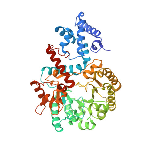
    Radical S-adenosyl-L-methionine (SAM) enzymes couple the reductive cleavage of SAM to radical-mediated transformations that have proven to be quite broad in scope. DesII is one such enzyme from the biosynthetic pathway of TDP-desosamine where it catalyzes a radical-mediated deamination. Previous studies have suggested that this reaction proceeds via direct elimination of ammonia from an α-hydroxyalkyl radical or its conjugate base (i.e., a ketyl radical) rather than 1,2-migration of the amino group to form a carbinolamine radical intermediate. However, without a crystal structure, the active site features responsible for this chemistry have remained largely unknown. The crystallographic studies described herein help to fill this gap by providing a structural description of the DesII active site. Computational analyses based on the solved crystal structure are consistent with direct elimination and indicate that an active site glutamate residue likely serves as a general base to promote deprotonation of the α-hydroxyalkyl radical intermediate and elimination of the ammonia group.


  • Organizational Affiliation
    • Shaanxi Key Laboratory of Natural Products & Chemical Biology, College of Chemistry & Pharmacy, Northwest A&F University, Yangling, Shaanxi, 712100, China.

Macromolecule Content 

  • Total Structure Weight: 220.42 kDa 
  • Atom Count: 15,451 
  • Modeled Residue Count: 1,902 
  • Deposited Residue Count: 1,972 
  • Unique protein chains: 1

Macromolecules

Find similar proteins by:|  3D Structure
Entity ID: 1
MoleculeChains  Sequence LengthOrganismDetailsImage
Radical S-Adenosyl-L-methionine Enzyme DesII
A, B, C, D
493Homo sapiensMutation(s): 0 
EC: 1.1.99 (UniProt), 4.3.1.30 (UniProt)
UniProt
Find proteins for Q9ZGH1 (Streptomyces venezuelae)
Explore Q9ZGH1 
Go to UniProtKB:  Q9ZGH1
Entity Groups
Sequence Clusters30% Identity50% Identity70% Identity90% Identity95% Identity100% Identity
UniProt GroupQ9ZGH1
Sequence Annotations
Expand
Reference Sequence

Small Molecules

Ligands 6 Unique
IDChains Name / Formula / InChI Key2D Diagram3D Interactions
SAM

Query on SAM


Download:Ideal Coordinates CCD File
F [auth A],
M [auth C]
S-ADENOSYLMETHIONINE
C15 H22 N6 O5 S
MEFKEPWMEQBLKI-FCKMPRQPSA-N
SF4

Query on SF4


Download:Ideal Coordinates CCD File
E [auth A],
H [auth B],
L [auth C],
P [auth D]
IRON/SULFUR CLUSTER
Fe4 S4
LJBDFODJNLIPKO-UHFFFAOYSA-N
MET
(Subject of Investigation/LOI)

Query on MET


Download:Ideal Coordinates CCD File
G [auth B],
O [auth D]
METHIONINE
C5 H11 N O2 S
FFEARJCKVFRZRR-BYPYZUCNSA-N
SO4

Query on SO4


Download:Ideal Coordinates CCD File
I [auth B],
N [auth C]
SULFATE ION
O4 S
QAOWNCQODCNURD-UHFFFAOYSA-L
GOL

Query on GOL


Download:Ideal Coordinates CCD File
J [auth B],
K [auth B],
R [auth D],
S [auth D]
GLYCEROL
C3 H8 O3
PEDCQBHIVMGVHV-UHFFFAOYSA-N
NA

Query on NA


Download:Ideal Coordinates CCD File
Q [auth D]SODIUM ION
Na
FKNQFGJONOIPTF-UHFFFAOYSA-N

Experimental Data & Validation

Experimental Data

  • Method: X-RAY DIFFRACTION
  • Resolution: 2.33 Å
  • R-Value Free:  0.235 (Depositor), 0.237 (DCC) 
  • R-Value Work:  0.204 (Depositor), 0.206 (DCC) 
  • R-Value Observed: 0.206 (Depositor) 
Space Group: P 1 21 1
Unit Cell:
Length ( Å )Angle ( ˚ )
a = 69.186α = 90
b = 140.79β = 96.279
c = 116.021γ = 90
Software Package:
Software NamePurpose
PHENIXrefinement
XDSdata reduction
Aimlessdata scaling
PHASERphasing

Structure Validation

View Full Validation Report


Ligand Structure Quality Assessment 


Entry History 

& Funding Information

Deposition Data


Funding OrganizationLocationGrant Number
Not funded--

Revision History  (Full details and data files)

  • Version 1.0: 2024-07-10
    Type: Initial release
  • Version 1.1: 2024-10-09
    Changes: Database references, Structure summary
  • Version 1.2: 2024-10-16
    Changes: Database references