8FE2 | pdb_00008fe2

Structure of J-PKAc chimera complexed with Aplithianine A


Experimental Data Snapshot

  • Method: X-RAY DIFFRACTION
  • Resolution: 2.34 Å
  • R-Value Free: 
    0.251 (Depositor), 0.251 (DCC) 
  • R-Value Work: 
    0.196 (Depositor), 0.196 (DCC) 
  • R-Value Observed: 
    0.199 (Depositor) 

Starting Model: experimental
View more details

wwPDB Validation 3D Report Full Report

Validation slider image for 8FE2

Ligand Structure Quality Assessment 


This is version 1.2 of the entry. See complete history

Literature

Discovery and Synthesis of a Naturally Derived Protein Kinase Inhibitor that Selectively Inhibits Distinct Classes of Serine/Threonine Kinases.

Du, L.Wilson, B.A.P.Li, N.Shah, R.Dalilian, M.Wang, D.Smith, E.A.Wamiru, A.Goncharova, E.I.Zhang, P.O'Keefe, B.R.

(2023) J Nat Prod 86: 2283-2293

  • DOI: https://doi.org/10.1021/acs.jnatprod.3c00394
  • Primary Citation Related Structures: 
    8FE2, 8FE5, 8FEC

  • PubMed Abstract: 

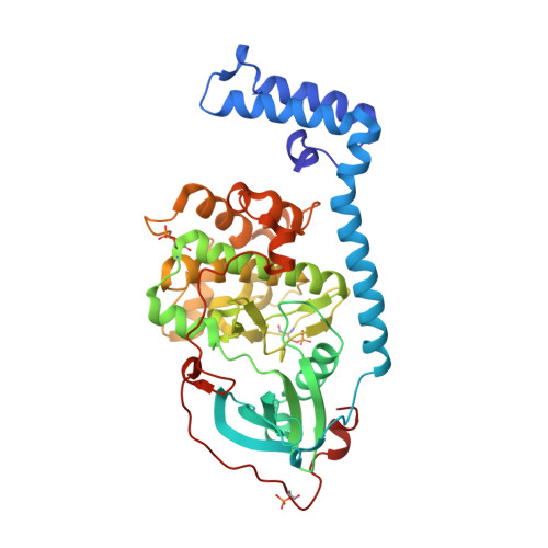
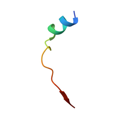
    The DNAJB1-PRKACA oncogenic gene fusion results in an active kinase enzyme, J-PKAcα, that has been identified as an attractive antitumor target for fibrolamellar hepatocellular carcinoma (FLHCC). A high-throughput assay was used to identify inhibitors of J-PKAcα catalytic activity by screening the NCI Program for Natural Product Discovery (NPNPD) prefractionated natural product library. Purification of the active agent from a single fraction of an Aplidium sp. marine tunicate led to the discovery of two unprecedented alkaloids, aplithianines A ( 1 ) and B ( 2 ). Aplithianine A ( 1 ) showed potent inhibition against J-PKAcα with an IC 50 of ∼1 μM in the primary screening assay. In kinome screening, 1 inhibited wild-type PKA with an IC 50 of 84 nM. Further mechanistic studies including cocrystallization and X-ray diffraction experiments revealed that 1 inhibited PKAcα catalytic activity by competitively binding to the ATP pocket. Human kinome profiling of 1 against a panel of 370 kinases revealed potent inhibition of select serine/threonine kinases in the CLK and PKG families with IC 50 values in the range ∼11-90 nM. An efficient, four-step total synthesis of 1 has been accomplished, enabling further evaluation of aplithianines as biologically relevant kinase inhibitors.


  • Organizational Affiliation
    • Molecular Targets Program, Center for Cancer Research, National Cancer Institute, Frederick, Maryland 21702, United States.

Macromolecule Content 

  • Total Structure Weight: 100.24 kDa 
  • Atom Count: 7,057 
  • Modeled Residue Count: 842 
  • Deposited Residue Count: 850 
  • Unique protein chains: 2

Macromolecules

Find similar proteins by:|  3D Structure
Entity ID: 1
MoleculeChains  Sequence LengthOrganismDetailsImage
DnaJ homolog subfamily B member 1, cAMP-dependent protein kinase catalytic subunit alpha
A, B
405Homo sapiensMutation(s): 0 
Gene Names: DNAJB1DNAJ1HDJ1HSPF1PRKACAPKACA
EC: 2.7.11.11
UniProt & NIH Common Fund Data Resources
Find proteins for P17612 (Homo sapiens)
Explore P17612 
Go to UniProtKB:  P17612
PHAROS:  P17612
GTEx:  ENSG00000072062 
Find proteins for P25685 (Homo sapiens)
Explore P25685 
Go to UniProtKB:  P25685
PHAROS:  P25685
GTEx:  ENSG00000132002 
Entity Groups
Sequence Clusters30% Identity50% Identity70% Identity90% Identity95% Identity100% Identity
UniProt GroupsP17612P25685
Sequence Annotations
Expand
Reference Sequence
Find similar proteins by:  Sequence   |   3D Structure  
Entity ID: 2
MoleculeChains  Sequence LengthOrganismDetailsImage
cAMP-dependent protein kinase inhibitor alphaC [auth I],
D [auth J]
20Homo sapiensMutation(s): 0 
UniProt & NIH Common Fund Data Resources
Find proteins for P61925 (Homo sapiens)
Explore P61925 
Go to UniProtKB:  P61925
PHAROS:  P61925
GTEx:  ENSG00000171033 
Entity Groups
Sequence Clusters30% Identity50% Identity70% Identity90% Identity95% Identity100% Identity
UniProt GroupP61925
Sequence Annotations
Expand
Reference Sequence

Small Molecules

Ligands 1 Unique
IDChains Name / Formula / InChI Key2D Diagram3D Interactions
XTI
(Subject of Investigation/LOI)

Query on XTI


Download:Ideal Coordinates CCD File
E [auth A],
F [auth B]
6-[(6M)-6-(1-methyl-1H-imidazol-5-yl)-2,3-dihydro-4H-1,4-thiazin-4-yl]-9H-purine
C13 H13 N7 S
IEDDDZFQZKDRBJ-UHFFFAOYSA-N
Modified Residues  2 Unique
IDChains TypeFormula2D DiagramParent
SEP
Query on SEP
A, B
L-PEPTIDE LINKINGC3 H8 N O6 PSER
TPO
Query on TPO
A, B
L-PEPTIDE LINKINGC4 H10 N O6 PTHR
Binding Affinity Annotations 
IDSourceBinding Affinity
XTI BindingDB:  8FE2 IC50: min: 84, max: 970 (nM) from 2 assay(s)

Experimental Data & Validation

Experimental Data

  • Method: X-RAY DIFFRACTION
  • Resolution: 2.34 Å
  • R-Value Free:  0.251 (Depositor), 0.251 (DCC) 
  • R-Value Work:  0.196 (Depositor), 0.196 (DCC) 
  • R-Value Observed: 0.199 (Depositor) 
Space Group: P 1 21 1
Unit Cell:
Length ( Å )Angle ( ˚ )
a = 50.633α = 90
b = 171.782β = 90.07
c = 61.481γ = 90
Software Package:
Software NamePurpose
XDSdata reduction
XDSdata scaling
PHASERphasing
PHENIXrefinement

Structure Validation

View Full Validation Report


Ligand Structure Quality Assessment 


Entry History 

& Funding Information

Deposition Data


Funding OrganizationLocationGrant Number
National Institutes of Health/National Cancer Institute (NIH/NCI)United States--

Revision History  (Full details and data files)

  • Version 1.0: 2023-10-18
    Type: Initial release
  • Version 1.1: 2024-05-01
    Changes: Database references
  • Version 1.2: 2024-11-20
    Changes: Structure summary