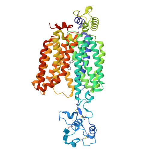
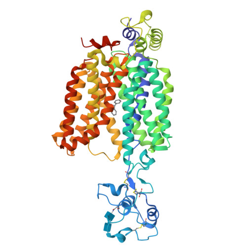
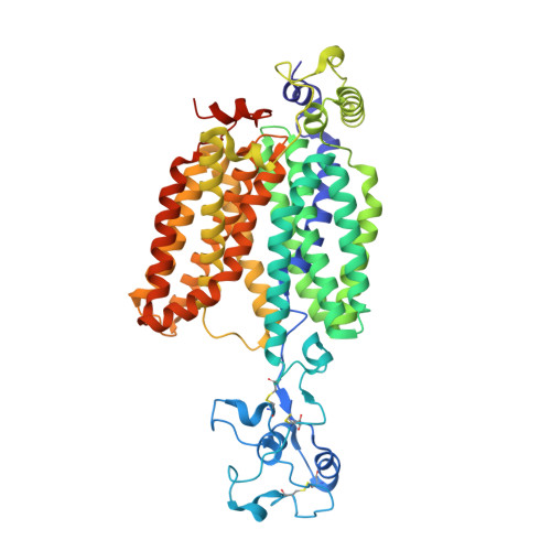
8ET7

Cryo-EM structure of the organic cation transporter 1 in complex with diphenhydramine


Experimental Data Snapshot

  • Method: ELECTRON MICROSCOPY
  • Resolution: 3.77 Å
  • Aggregation State: PARTICLE 
  • Reconstruction Method: SINGLE PARTICLE 

wwPDB Validation   3D Report Full Report


This is version 1.3 of the entry. See complete history


Literature

Molecular basis of polyspecific drug and xenobiotic recognition by OCT1 and OCT2.

Suo, Y.Wright, N.J.Guterres, H.Fedor, J.G.Butay, K.J.Borgnia, M.J.Im, W.Lee, S.Y.

(2023) Nat Struct Mol Biol 30: 1001-1011

  • DOI: https://doi.org/10.1038/s41594-023-01017-4
  • Primary Citation of Related Structures:  
    8ET6, 8ET7, 8ET8, 8ET9

  • PubMed Abstract: 

    A wide range of endogenous and xenobiotic organic ions require facilitated transport systems to cross the plasma membrane for their disposition. In mammals, organic cation transporter (OCT) subtypes 1 and 2 (OCT1 and OCT2, also known as SLC22A1 and SLC22A2, respectively) are polyspecific transporters responsible for the uptake and clearance of structurally diverse cationic compounds in the liver and kidneys, respectively. Notably, it is well established that human OCT1 and OCT2 play central roles in the pharmacokinetics and drug-drug interactions of many prescription medications, including metformin. Despite their importance, the basis of polyspecific cationic drug recognition and the alternating access mechanism for OCTs have remained a mystery. Here we present four cryo-electron microscopy structures of apo, substrate-bound and drug-bound OCT1 and OCT2 consensus variants, in outward-facing and outward-occluded states. Together with functional experiments, in silico docking and molecular dynamics simulations, these structures uncover general principles of organic cation recognition by OCTs and provide insights into extracellular gate occlusion. Our findings set the stage for a comprehensive structure-based understanding of OCT-mediated drug-drug interactions, which will prove critical in the preclinical evaluation of emerging therapeutics.


  • Organizational Affiliation

    Department of Biochemistry, Duke University School of Medicine, Durham, NC, USA.


Macromolecules
Find similar proteins by:  (by identity cutoff)  |  3D Structure
Entity ID: 1
MoleculeChains Sequence LengthOrganismDetailsImage
OCT1554Homo sapiensMutation(s): 0 
UniProt & NIH Common Fund Data Resources
Find proteins for O15245 (Homo sapiens)
Go to UniProtKB:  O15245
PHAROS:  O15245
GTEx:  ENSG00000175003 
Glycosylation
Glycosylation Sites: 1Go to GlyGen: O15245-1
Sequence Annotations
Expand
  • Reference Sequence
Oligosaccharides

Help

Entity ID: 2
MoleculeChains Length2D Diagram Glycosylation3D Interactions
2-acetamido-2-deoxy-beta-D-glucopyranose-(1-4)-2-acetamido-2-deoxy-beta-D-glucopyranose
B
2N-Glycosylation
Glycosylation Resources
GlyTouCan:  G42666HT
GlyCosmos:  G42666HT
GlyGen:  G42666HT
Small Molecules
Ligands 1 Unique
IDChains Name / Formula / InChI Key2D Diagram3D Interactions
2PM (Subject of Investigation/LOI)
Query on 2PM

Download Ideal Coordinates CCD File 
C [auth A]N-[2-(BENZHYDRYLOXY)ETHYL]-N,N-DIMETHYLAMINE
C17 H21 N O
ZZVUWRFHKOJYTH-UHFFFAOYSA-N
Experimental Data & Validation

Experimental Data

  • Method: ELECTRON MICROSCOPY
  • Resolution: 3.77 Å
  • Aggregation State: PARTICLE 
  • Reconstruction Method: SINGLE PARTICLE 
EM Software:
TaskSoftware PackageVersion
RECONSTRUCTIONcryoSPARC3.2
MODEL REFINEMENTPHENIX1.19

Structure Validation

View Full Validation Report


Entry History & Funding Information

Deposition Data


Funding OrganizationLocationGrant Number
National Institutes of Health/National Institute of General Medical Sciences (NIH/NIGMS)United StatesR01GM137421

Revision History  (Full details and data files)

  • Version 1.0: 2023-05-31
    Type: Initial release
  • Version 1.1: 2023-07-12
    Changes: Database references
  • Version 1.2: 2023-08-02
    Changes: Database references
  • Version 1.3: 2024-10-23
    Changes: Data collection, Structure summary