8D5K | pdb_00008d5k

The complex of Pre-mRNA-Processing Factor 19 (Prpf19) peptide KYLQVASHV Presented by H2-Kd


Experimental Data Snapshot

  • Method: X-RAY DIFFRACTION
  • Resolution: 2.07 Å
  • R-Value Free: 
    0.234 (Depositor), 0.230 (DCC) 
  • R-Value Work: 
    0.203 (Depositor), 0.210 (DCC) 

Starting Model: experimental
View more details

wwPDB Validation 3D Report Full Report

Validation slider image for 8D5K

This is version 1.3 of the entry. See complete history

Literature

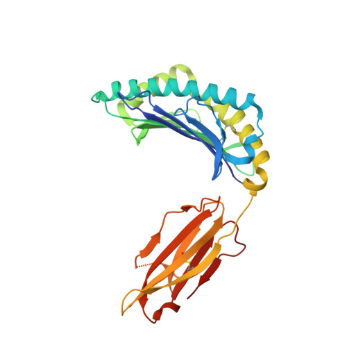
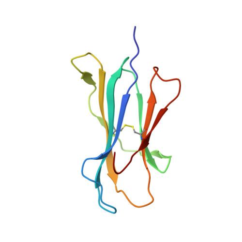
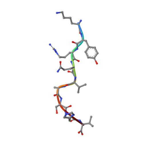
Structural and physical features that distinguish tumor-controlling from inactive cancer neoepitopes.

Custodio, J.M.Ayres, C.M.Rosales, T.J.Brambley, C.A.Arbuiso, A.G.Landau, L.M.Keller, G.L.J.Srivastava, P.K.Baker, B.M.

(2023) Proc Natl Acad Sci U S A 120: e2312057120-e2312057120

  • DOI: https://doi.org/10.1073/pnas.2312057120
  • Primary Citation Related Structures: 
    8D5E, 8D5F, 8D5K

  • PubMed Abstract: 

    Neoepitopes arising from amino acid substitutions due to single nucleotide polymorphisms are targets of T cell immune responses to cancer and are of significant interest in the development of cancer vaccines. However, understanding the characteristics of rare protective neoepitopes that truly control tumor growth has been a challenge, due to their scarcity as well as the challenge of verifying true, neoepitope-dependent tumor control in humans. Taking advantage of recent work in mouse models that circumvented these challenges, here, we compared the structural and physical properties of neoepitopes that range from fully protective to immunologically inactive. As neoepitopes are derived from self-peptides that can induce immune tolerance, we studied not only how the various neoepitopes differ from each other but also from their wild-type counterparts. We identified multiple features associated with protection, including features that describe how neoepitopes differ from self as well as features associated with recognition by diverse T cell receptor repertoires. We demonstrate both the promise and limitations of neoepitope structural analysis and predictive modeling and illustrate important aspects that can be incorporated into neoepitope prediction pipelines.


  • Organizational Affiliation
    • Department of Chemistry and Biochemistry and the Harper Cancer Research Institute, University of Notre Dame, Notre Dame, IN 46556.

Macromolecule Content 

  • Total Structure Weight: 45.04 kDa 
  • Atom Count: 3,194 
  • Modeled Residue Count: 382 
  • Deposited Residue Count: 384 
  • Unique protein chains: 3

Macromolecules

Find similar proteins by:|  3D Structure
Entity ID: 1
MoleculeChains  Sequence LengthOrganismDetailsImage
H-2 class I histocompatibility antigen, K-D alpha chain275Mus musculusMutation(s): 0 
Gene Names: H2-K1H2-K
UniProt
Find proteins for P01902 (Mus musculus)
Explore P01902 
Go to UniProtKB:  P01902
Entity Groups
Sequence Clusters30% Identity50% Identity70% Identity90% Identity95% Identity100% Identity
UniProt GroupP01902
Sequence Annotations
Expand
Reference Sequence
Find similar proteins by:|  3D Structure
Entity ID: 2
MoleculeChains  Sequence LengthOrganismDetailsImage
Beta-2-microglobulin100Mus musculusMutation(s): 0 
Gene Names: B2m
UniProt & NIH Common Fund Data Resources
Find proteins for P01887 (Mus musculus)
Explore P01887 
Go to UniProtKB:  P01887
IMPC:  MGI:88127
Entity Groups
Sequence Clusters30% Identity50% Identity70% Identity90% Identity95% Identity100% Identity
UniProt GroupP01887
Sequence Annotations
Expand
Reference Sequence
Find similar proteins by:  Sequence   |   3D Structure  
Entity ID: 3
MoleculeChains  Sequence LengthOrganismDetailsImage
Pre-mRNA-processing factor 199Mus musculusMutation(s): 0 
EC: 2.3.2.27
UniProt
Find proteins for Q99KP6 (Mus musculus)
Explore Q99KP6 
Go to UniProtKB:  Q99KP6
Entity Groups
UniProt GroupQ99KP6
Sequence Annotations
Expand
Reference Sequence

Experimental Data & Validation

Experimental Data

  • Method: X-RAY DIFFRACTION
  • Resolution: 2.07 Å
  • R-Value Free:  0.234 (Depositor), 0.230 (DCC) 
  • R-Value Work:  0.203 (Depositor), 0.210 (DCC) 
Space Group: C 1 2 1
Unit Cell:
Length ( Å )Angle ( ˚ )
a = 115.37α = 90
b = 66.2β = 92.42
c = 56.79γ = 90
Software Package:
Software NamePurpose
REFMACrefinement
REFMACrefinement
BUCCANEERmodel building
SCALEPACKdata scaling
PHASERphasing

Structure Validation

View Full Validation Report


Entry History 

& Funding Information

Deposition Data


Funding OrganizationLocationGrant Number
National Institutes of Health/National Institute of General Medical Sciences (NIH/NIGMS)United StatesR35GM118166

Revision History  (Full details and data files)

  • Version 1.0: 2022-07-06
    Type: Initial release
  • Version 1.1: 2023-10-18
    Changes: Data collection, Refinement description
  • Version 1.2: 2024-01-17
    Changes: Database references
  • Version 1.3: 2024-10-16
    Changes: Structure summary