8COL

Crystal structure of Rhizobium etli constitutive L-asparaginase ReAIV (orthorombic form R4oP-2)


Experimental Data Snapshot

  • Method: X-RAY DIFFRACTION
  • Resolution: 2.00 Å
  • R-Value Free: 0.234 
  • R-Value Work: 0.177 

wwPDB Validation   3D Report Full Report


This is version 1.0 of the entry. See complete history


Literature

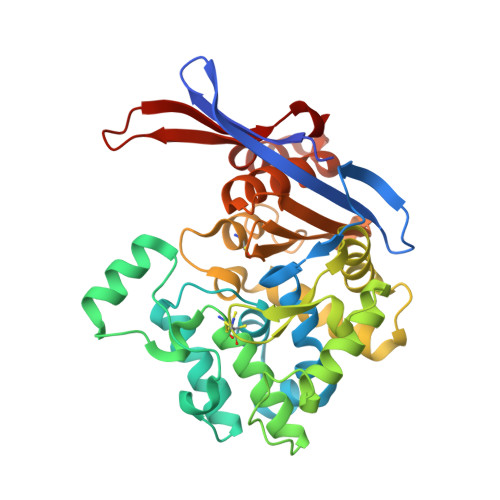
Rhizobium etli has two L-asparaginases with low sequence identity but similar structure and catalytic center.

Loch, J.I.Worsztynowicz, P.Sliwiak, J.Grzechowiak, M.Imiolczyk, B.Pokrywka, K.Chwastyk, M.Gilski, M.Jaskolski, M.

(2023) Acta Crystallogr D Struct Biol 79: 775-791

  • DOI: https://doi.org/10.1107/S2059798323005648
  • Primary Citation of Related Structures:  
    8CLY, 8CLZ, 8COL, 8ORI, 8OSW

  • PubMed Abstract: 

    The genome of Rhizobium etli, a nitrogen-fixing bacterial symbiont of legume plants, encodes two L-asparaginases, ReAIV and ReAV, that have no similarity to the well characterized enzymes of class 1 (bacterial type) and class 2 (plant type). It has been hypothesized that ReAIV and ReAV might belong to the same structural class 3 despite their low level of sequence identity. When the crystal structure of the inducible and thermolabile protein ReAV was solved, this hypothesis gained a stronger footing because the key residues of ReAV are also present in the sequence of the constitutive and thermostable ReAIV protein. High-resolution crystal structures of ReAIV now confirm that it is a class 3 L-asparaginase that is structurally similar to ReAV but with important differences. The most striking differences concern the peculiar hydration patterns of the two proteins, the presence of three internal cavities in ReAIV and the behavior of the zinc-binding site. ReAIV has a high pH optimum (9-11) and a substrate affinity of ∼1.3 mM at pH 9.0. These parameters are not suitable for the direct application of ReAIV as an antileukemic drug, although its thermal stability and lack of glutaminase activity would be of considerable advantage. The five crystal structures of ReAIV presented in this work allow a possible enzymatic scenario to be postulated in which the zinc ion coordinated in the active site is a dispensable element. The catalytic nucleophile seems to be Ser47, which is part of two Ser-Lys tandems in the active site. The structures of ReAIV presented here may provide a basis for future enzyme-engineering experiments to improve the kinetic parameters for medicinal applications.


  • Organizational Affiliation

    Department of Crystal Chemistry and Crystal Physics, Faculty of Chemistry, Jagiellonian University, Krakow, Poland.


Macromolecules
Find similar proteins by:  (by identity cutoff)  |  3D Structure
Entity ID: 1
MoleculeChains Sequence LengthOrganismDetailsImage
Putative L-asparaginase II proteinA [auth AAA],
B [auth BBB],
C [auth CCC],
D [auth DDD]
341Rhizobium etliMutation(s): 0 
Gene Names: RHE_CH01144
UniProt
Find proteins for Q2KB35 (Rhizobium etli (strain ATCC 51251 / DSM 11541 / JCM 21823 / NBRC 15573 / CFN 42))
Explore Q2KB35 
Go to UniProtKB:  Q2KB35
Entity Groups  
Sequence Clusters30% Identity50% Identity70% Identity90% Identity95% Identity100% Identity
UniProt GroupQ2KB35
Sequence Annotations
Expand
  • Reference Sequence
Small Molecules
Ligands 4 Unique
IDChains Name / Formula / InChI Key2D Diagram3D Interactions
ZN
Query on ZN

Download Ideal Coordinates CCD File 
E [auth AAA],
K [auth BBB],
N [auth CCC],
R [auth DDD]
ZINC ION
Zn
PTFCDOFLOPIGGS-UHFFFAOYSA-N
EDO
Query on EDO

Download Ideal Coordinates CCD File 
H [auth AAA],
I [auth AAA],
S [auth DDD],
W [auth DDD]
1,2-ETHANEDIOL
C2 H6 O2
LYCAIKOWRPUZTN-UHFFFAOYSA-N
CL
Query on CL

Download Ideal Coordinates CCD File 
F [auth AAA]
G [auth AAA]
J [auth AAA]
L [auth BBB]
M [auth BBB]
F [auth AAA],
G [auth AAA],
J [auth AAA],
L [auth BBB],
M [auth BBB],
O [auth CCC],
Q [auth CCC],
T [auth DDD],
U [auth DDD]
CHLORIDE ION
Cl
VEXZGXHMUGYJMC-UHFFFAOYSA-M
MG
Query on MG

Download Ideal Coordinates CCD File 
P [auth CCC],
V [auth DDD]
MAGNESIUM ION
Mg
JLVVSXFLKOJNIY-UHFFFAOYSA-N
Modified Residues  1 Unique
IDChains TypeFormula2D DiagramParent
CSO
Query on CSO
A [auth AAA],
B [auth BBB],
C [auth CCC],
D [auth DDD]
L-PEPTIDE LINKINGC3 H7 N O3 SCYS
Experimental Data & Validation

Experimental Data

  • Method: X-RAY DIFFRACTION
  • Resolution: 2.00 Å
  • R-Value Free: 0.234 
  • R-Value Work: 0.177 
  • Space Group: P 21 21 21
Unit Cell:
Length ( Å )Angle ( ˚ )
a = 83.101α = 90
b = 89.749β = 90
c = 169.197γ = 90
Software Package:
Software NamePurpose
REFMACrefinement
XDSdata reduction
XDSdata scaling
PHASERphasing

Structure Validation

View Full Validation Report


Entry History & Funding Information

Deposition Data


Funding OrganizationLocationGrant Number
Polish National Science CentrePoland2020/37/B/NZ1/03250

Revision History  (Full details and data files)

  • Version 1.0: 2023-08-09
    Type: Initial release