8BOV | pdb_00008bov

X-ray structure of the adduct formed upon reaction of the five-coordinate Pt(II) complex, 1-Me,Me, with HEWL at pH 7.5


Experimental Data Snapshot

  • Method: X-RAY DIFFRACTION
  • Resolution: 1.25 Å
  • R-Value Free: 
    0.218 (Depositor), 0.242 (DCC) 
  • R-Value Work: 
    0.182 (Depositor) 

Starting Model: experimental
View more details

wwPDB Validation 3D Report Full Report

Validation slider image for 8BOV

Ligand Structure Quality Assessment 


This is version 1.2 of the entry. See complete history

Literature

Impact of Hydrophobic Chains in Five-Coordinate Glucoconjugate Pt(II) Anticancer Agents.

Annunziata, A.Imbimbo, P.Cucciolito, M.E.Ferraro, G.Langellotti, V.Marano, A.Melchiorre, M.Tito, G.Trifuoggi, M.Monti, D.M.Merlino, A.Ruffo, F.

(2023) Int J Mol Sci 24

  • DOI: https://doi.org/10.3390/ijms24032369
  • Primary Citation Related Structures: 
    8BOV, 8BOY

  • PubMed Abstract: 

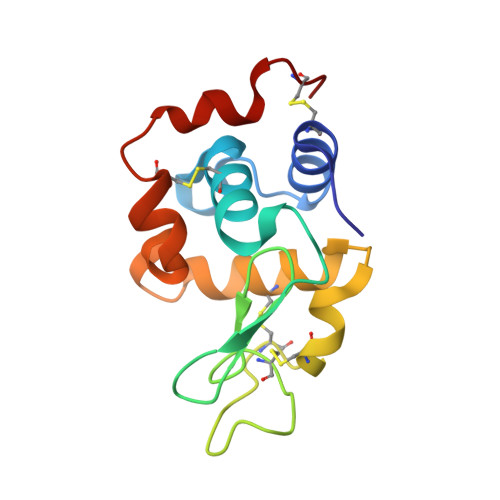
    This study describes new platinum(II) cationic five-coordinate complexes ( 1-R,R' ) of the formula [PtR(NHC)(dmphen)(ethene)]CF 3 SO 3 (dmphen = 2,9-dimethyl-1,10-phenanthroline), containing in their axial positions an alkyl group R (methyl or octyl) and an imidazole-based NHC-carbene ligand with a substituent R' of variable length (methyl or octyl) on one nitrogen atom. The Pt-carbene bond is stable both in DMSO and in aqueous solvents. In DMSO, a gradual substitution of dmphen and ethene is observed, with the formation of a square planar solvated species. Octanol/water partitioning studies have revealed the order of hydrophobicity of the complexes ( 1-Oct,Me > 1-Oct,Oct > 1-Me,Oct > 1-Me,Me ). Their biological activity was investigated against two pairs of cancer and non-cancer cell lines. The tested drugs were internalized in cancer cells and able to activate the apoptotic pathway. The reactivity of 1-Me,Me with DNA and protein model systems was also studied using UV-vis absorption spectroscopy, fluorescence, and X-ray crystallography. The compound binds DNA and interacts in various ways with the model protein lysozyme. Remarkably, structural data revealed that the complex can bind lysozyme via non-covalent interactions, retaining its five-coordinate geometry.


  • Organizational Affiliation
    • Institute Parisien de Chimie Moléculaire, Campus Pierre et Marie Curie, Sorbonne Université, 4 Place Jussieu, 75005 Paris, France.

Macromolecule Content 

  • Total Structure Weight: 15.37 kDa 
  • Atom Count: 1,254 
  • Modeled Residue Count: 129 
  • Deposited Residue Count: 129 
  • Unique protein chains: 1

Macromolecules

Find similar proteins by:|  3D Structure
Entity ID: 1
MoleculeChains  Sequence LengthOrganismDetailsImage
LysozymeA [auth AAA]129Gallus gallusMutation(s): 0 
EC: 3.2.1.17
UniProt
Find proteins for P00698 (Gallus gallus)
Explore P00698 
Go to UniProtKB:  P00698
Entity Groups
Sequence Clusters30% Identity50% Identity70% Identity90% Identity95% Identity100% Identity
UniProt GroupP00698
Sequence Annotations
Expand
Reference Sequence

Small Molecules

Ligands 4 Unique
IDChains Name / Formula / InChI Key2D Diagram3D Interactions
R0I
(Subject of Investigation/LOI)

Query on R0I


Download:Ideal Coordinates CCD File
E [auth AAA]1-[1,3-dimethyl-4-(1~{H}-1,2,3-triazol-5-yl)imidazol-1-ium-2-yl]-1,2',11'-trimethyl-spiro[1$l^{6}-platinacycloprop-2-ene-1,15'-1,12-diaza-15$l^{6}-platinatetracyclo[10.2.1.0^{5,14}.0^{8,13}]pentadeca-2,4,6,8,10,13-hexaene]
C24 H26 N7 Pt
DGCRSFURYLTMIA-UHFFFAOYSA-N
EPE

Query on EPE


Download:Ideal Coordinates CCD File
B [auth AAA]4-(2-HYDROXYETHYL)-1-PIPERAZINE ETHANESULFONIC ACID
C8 H18 N2 O4 S
JKMHFZQWWAIEOD-UHFFFAOYSA-N
DMS

Query on DMS


Download:Ideal Coordinates CCD File
F [auth AAA]DIMETHYL SULFOXIDE
C2 H6 O S
IAZDPXIOMUYVGZ-UHFFFAOYSA-N
ACT

Query on ACT


Download:Ideal Coordinates CCD File
C [auth AAA],
D [auth AAA]
ACETATE ION
C2 H3 O2
QTBSBXVTEAMEQO-UHFFFAOYSA-M

Experimental Data & Validation

Experimental Data

  • Method: X-RAY DIFFRACTION
  • Resolution: 1.25 Å
  • R-Value Free:  0.218 (Depositor), 0.242 (DCC) 
  • R-Value Work:  0.182 (Depositor) 
Space Group: P 43 21 2
Unit Cell:
Length ( Å )Angle ( ˚ )
a = 78.58α = 90
b = 78.58β = 90
c = 37.05γ = 90
Software Package:
Software NamePurpose
REFMACrefinement
autoPROCdata reduction
autoPROCdata scaling
PHASERphasing

Structure Validation

View Full Validation Report


Ligand Structure Quality Assessment 


Entry History 

& Funding Information

Deposition Data


Funding OrganizationLocationGrant Number
Not funded--

Revision History  (Full details and data files)

  • Version 1.0: 2023-02-22
    Type: Initial release
  • Version 1.1: 2024-02-07
    Changes: Data collection, Refinement description
  • Version 1.2: 2024-11-06
    Changes: Structure summary