8BFD | pdb_00008bfd

Racemic structure of PK-7 (310HD-U2U5)


Experimental Data Snapshot

  • Method: X-RAY DIFFRACTION
  • Resolution: 2.00 Å
  • R-Value Free: 
    0.256 (Depositor), 0.270 (DCC) 
  • R-Value Work: 
    0.203 (Depositor), 0.204 (DCC) 
  • R-Value Observed: 
    0.209 (Depositor) 

wwPDB Validation 3D Report Full Report

Validation slider image for 8BFD

This is version 1.0 of the entry. See complete history

Literature

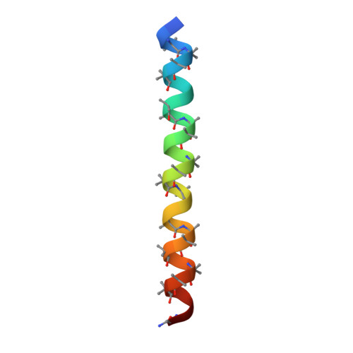
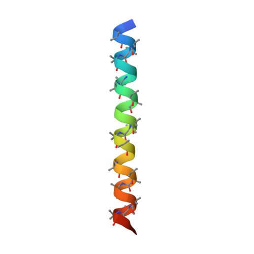
De novo design of discrete, stable 3 10 -helix peptide assemblies.

Kumar, P.Paterson, N.G.Clayden, J.Woolfson, D.N.

(2022) Nature 607: 387-392

  • DOI: https://doi.org/10.1038/s41586-022-04868-x
  • Primary Citation Related Structures: 
    7QDI, 7QDJ, 7QDK, 8BFD, 8BFE

  • PubMed Abstract: 

    The α-helix is pre-eminent in structural biology 1 and widely exploited in protein folding 2 , design 3 and engineering 4 . Although other helical peptide conformations do exist near to the α-helical region of conformational space-namely, 3 10 -helices and π-helices 5 -these occur much less frequently in protein structures. Less favourable internal energies and reduced tendencies to pack into higher-order structures mean that 3 10 -helices rarely exceed six residues in length in natural proteins, and that they tend not to form normal supersecondary, tertiary or quaternary interactions. Here we show that despite their absence in nature, synthetic peptide assemblies can be built from 3 10 -helices. We report the rational design, solution-phase characterization and an X-ray crystal structure for water-soluble bundles of 3 10 -helices with consolidated hydrophobic cores. The design uses six-residue repeats informed by analysing 3 10 -helical conformations in known protein structures, and incorporates α-aminoisobutyric acid residues. Design iterations reveal a tipping point between α-helical and 3 10 -helical folding, and identify features required for stabilizing assemblies of 3 10 -helices. This work provides principles and rules to open opportunities for designing into this hitherto unexplored region of protein-structure space.


  • Organizational Affiliation
    • School of Chemistry, University of Bristol, Bristol, UK. prasun.kumar@bristol.ac.uk.

Macromolecule Content 

  • Total Structure Weight: 6.44 kDa 
  • Atom Count: 475 
  • Modeled Residue Count: 64 
  • Deposited Residue Count: 66 
  • Unique protein chains: 2

Macromolecules

Find similar proteins by:|  3D Structure
Entity ID: 1
MoleculeChains  Sequence LengthOrganismDetailsImage
310HD-U2U533synthetic constructMutation(s): 0 
Find similar proteins by:|  3D Structure
Entity ID: 2
MoleculeChains  Sequence LengthOrganismDetailsImage
D-310HD-U2U533synthetic constructMutation(s): 0 

Experimental Data & Validation

Experimental Data

  • Method: X-RAY DIFFRACTION
  • Resolution: 2.00 Å
  • R-Value Free:  0.256 (Depositor), 0.270 (DCC) 
  • R-Value Work:  0.203 (Depositor), 0.204 (DCC) 
  • R-Value Observed: 0.209 (Depositor) 
Space Group: P 1
Unit Cell:
Length ( Å )Angle ( ˚ )
a = 21.23α = 105.85
b = 21.905β = 112.44
c = 26.521γ = 95.26
Software Package:
Software NamePurpose
Aimlessdata scaling
REFMACrefinement
PDB_EXTRACTdata extraction
xia2data reduction
Arcimboldophasing

Structure Validation

View Full Validation Report


Entry History 

& Funding Information

Deposition Data


Funding OrganizationLocationGrant Number
Biotechnology and Biological Sciences Research Council (BBSRC)United KingdomBB/L01386X/1
Biotechnology and Biological Sciences Research Council (BBSRC)United KingdomBB/R00661X/1

Revision History  (Full details and data files)

  • Version 1.0: 2022-11-23
    Type: Initial release