8A18

1.63 A resolution hydroquinone inhibited Sporosarcina pasteurii urease


Experimental Data Snapshot

  • Method: X-RAY DIFFRACTION
  • Resolution: 1.63 Å
  • R-Value Free: 0.158 
  • R-Value Work: 0.128 

Starting Model: experimental
View more details

wwPDB Validation   3D Report Full Report


Ligand Structure Quality Assessment 

Created with Raphaël 2.3.0Worse 01 BetterLigand structure goodness of fit to experimental dataBest fitted HQEClick on this verticalbar to view details

This is version 1.2 of the entry. See complete history


Literature

Inhibition of Urease by Hydroquinones: A Structural and Kinetic Study.

Mazzei, L.Cianci, M.Ciurli, S.

(2022) Chemistry 28: e202201770-e202201770

  • DOI: https://doi.org/10.1002/chem.202201770
  • Primary Citation of Related Structures:  
    8A18

  • PubMed Abstract: 

    Hydroquinones are a class of organic compounds abundant in nature that result from the full reduction of the corresponding quinones. Quinones are known to efficiently inhibit urease, a Ni II -containing enzyme that catalyzes the hydrolysis of urea to yield ammonia and carbonate and acts as a virulence factor of several human pathogens, in addition to decreasing the efficiency of soil organic nitrogen fertilization. Here, we report the molecular characterization of the inhibition of urease from Sporosarcina pasteurii (SPU) and Canavalia ensiformis (jack bean, JBU) by 1,4-hydroquinone (HQ) and its methyl and tert-butyl derivatives. The 1.63-Å resolution X-ray crystal structure of the SPU-HQ complex discloses that HQ covalently binds to the thiol group of αCys322, a key residue located on a mobile protein flap directly involved in the catalytic mechanism. Inhibition kinetic data obtained for the three compounds on JBU reveals the occurrence of an irreversible inactivation process that involves a radical-based autocatalytic mechanism.


  • Organizational Affiliation

    Laboratory of Bioinorganic Chemistry Department of Pharmacy and Biotechnology (FaBiT), University of Bologna, Viale Giuseppe Fanin 40, 40127, Bologna, Italy.


Macromolecules
Find similar proteins by:  (by identity cutoff)  |  3D Structure
Entity ID: 1
MoleculeChains Sequence LengthOrganismDetailsImage
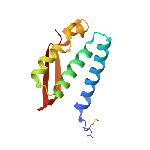
Urease subunit gammaA [auth AAA]100Sporosarcina pasteuriiMutation(s): 2 
EC: 3.5.1.5
UniProt
Find proteins for P41022 (Sporosarcina pasteurii)
Explore P41022 
Go to UniProtKB:  P41022
Entity Groups  
Sequence Clusters30% Identity50% Identity70% Identity90% Identity95% Identity100% Identity
UniProt GroupP41022
Sequence Annotations
Expand
  • Reference Sequence
Find similar proteins by:  (by identity cutoff)  |  3D Structure
Entity ID: 2
MoleculeChains Sequence LengthOrganismDetailsImage
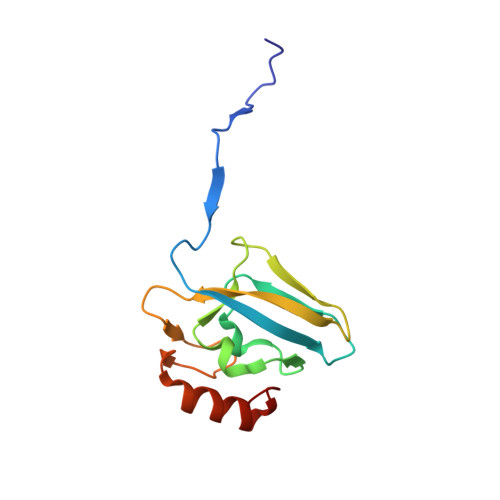
Urease subunit betaB [auth BBB]122Sporosarcina pasteuriiMutation(s): 0 
EC: 3.5.1.5
UniProt
Find proteins for P41021 (Sporosarcina pasteurii)
Explore P41021 
Go to UniProtKB:  P41021
Entity Groups  
Sequence Clusters30% Identity50% Identity70% Identity90% Identity95% Identity100% Identity
UniProt GroupP41021
Sequence Annotations
Expand
  • Reference Sequence
Find similar proteins by:  (by identity cutoff)  |  3D Structure
Entity ID: 3
MoleculeChains Sequence LengthOrganismDetailsImage
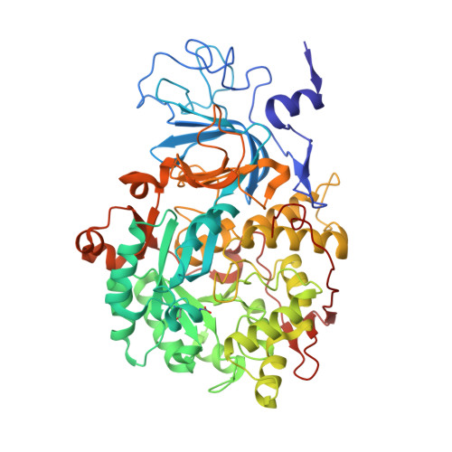
Urease subunit alphaC [auth CCC]570Sporosarcina pasteuriiMutation(s): 0 
EC: 3.5.1.5
UniProt
Find proteins for P41020 (Sporosarcina pasteurii)
Explore P41020 
Go to UniProtKB:  P41020
Entity Groups  
Sequence Clusters30% Identity50% Identity70% Identity90% Identity95% Identity100% Identity
UniProt GroupP41020
Sequence Annotations
Expand
  • Reference Sequence
Small Molecules
Ligands 5 Unique
IDChains Name / Formula / InChI Key2D Diagram3D Interactions
HQE (Subject of Investigation/LOI)
Query on HQE

Download Ideal Coordinates CCD File 
JA [auth CCC],
KA [auth CCC]
benzene-1,4-diol
C6 H6 O2
QIGBRXMKCJKVMJ-UHFFFAOYSA-N
SO4
Query on SO4

Download Ideal Coordinates CCD File 
I [auth AAA]
J [auth AAA]
K [auth AAA]
L [auth BBB]
LA [auth CCC]
SULFATE ION
O4 S
QAOWNCQODCNURD-UHFFFAOYSA-L
EDO
Query on EDO

Download Ideal Coordinates CCD File 
AA [auth CCC]
BA [auth CCC]
CA [auth CCC]
D [auth AAA]
DA [auth CCC]
1,2-ETHANEDIOL
C2 H6 O2
LYCAIKOWRPUZTN-UHFFFAOYSA-N
NI
Query on NI

Download Ideal Coordinates CCD File 
R [auth CCC],
S [auth CCC]
NICKEL (II) ION
Ni
VEQPNABPJHWNSG-UHFFFAOYSA-N
OH
Query on OH

Download Ideal Coordinates CCD File 
T [auth CCC]HYDROXIDE ION
H O
XLYOFNOQVPJJNP-UHFFFAOYSA-M
Modified Residues  2 Unique
IDChains TypeFormula2D DiagramParent
CXM
Query on CXM
A [auth AAA]L-PEPTIDE LINKINGC6 H11 N O4 SMET
KCX
Query on KCX
C [auth CCC]L-PEPTIDE LINKINGC7 H14 N2 O4LYS
Experimental Data & Validation

Experimental Data

  • Method: X-RAY DIFFRACTION
  • Resolution: 1.63 Å
  • R-Value Free: 0.158 
  • R-Value Work: 0.128 
  • Space Group: P 63 2 2
Unit Cell:
Length ( Å )Angle ( ˚ )
a = 131.666α = 90
b = 131.666β = 90
c = 189.129γ = 120
Software Package:
Software NamePurpose
REFMACrefinement
XDSdata reduction
Aimlessdata scaling
REFMACphasing

Structure Validation

View Full Validation Report


Ligand Structure Quality Assessment 

Created with Raphaël 2.3.0Worse 01 BetterLigand structure goodness of fit to experimental dataBest fitted HQEClick on this verticalbar to view details

Entry History & Funding Information

Deposition Data


Funding OrganizationLocationGrant Number
Not funded--

Revision History  (Full details and data files)

  • Version 1.0: 2023-06-14
    Type: Initial release
  • Version 1.1: 2023-12-27
    Changes: Data collection, Database references
  • Version 1.2: 2024-02-07
    Changes: Refinement description