Micro-electron diffraction structure of the aggregation-driving N terminus of Drosophila neuronal protein Orb2A reveals amyloid-like beta-sheets.
Bowler, J.T., Sawaya, M.R., Boyer, D.R., Cascio, D., Bali, M., Eisenberg, D.S.(2022) J Biological Chem 298: 102396-102396
- PubMed: 35988647 Search on PubMedSearch on PubMed Central
- DOI: https://doi.org/10.1016/j.jbc.2022.102396
- Primary Citation Related Structures:
7SXN - PubMed Abstract:
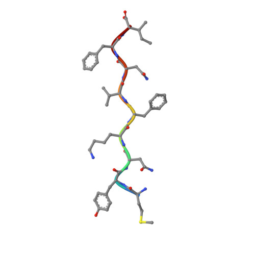
Amyloid protein aggregation is commonly associated with progressive neurodegenerative diseases, however not all amyloid fibrils are pathogenic. The neuronal cytoplasmic polyadenylation element binding protein is a regulator of synaptic mRNA translation and has been shown to form functional amyloid aggregates that stabilize long-term memory. In adult Drosophila neurons, the cytoplasmic polyadenylation element binding homolog Orb2 is expressed as 2 isoforms, of which the Orb2B isoform is far more abundant, but the rarer Orb2A isoform is required to initiate Orb2 aggregation. The N terminus is a distinctive feature of the Orb2A isoform and is critical for its aggregation. Intriguingly, replacement of phenylalanine in the fifth position of Orb2A with tyrosine (F5Y) in Drosophila impairs stabilization of long-term memory. The structure of endogenous Orb2B fibers was recently determined by cryo-EM, but the structure adopted by fibrillar Orb2A is less certain. Here we use micro-electron diffraction to determine the structure of the first 9 N-terminal residues of Orb2A, at a resolution of 1.05 Å. We find that this segment (which we term M9I) forms an amyloid-like array of parallel in-register β-sheets, which interact through side chain interdigitation of aromatic and hydrophobic residues. Our structure provides an explanation for the decreased aggregation observed for the F5Y mutant and offers a hypothesis for how the addition of a single atom (the tyrosyl oxygen) affects long-term memory. We also propose a structural model of Orb2A that integrates our structure of the M9I segment with the published Orb2B cryo-EM structure.
- Department of Biological Chemistry, UCLA-DOE Institute, Howard Hughes Medical Institute, and Molecular Biology Institute, UCLA, Los Angeles, California, USA. Electronic address: jbowler@ucla.edu.
Organizational Affiliation: