7ZJP

Optimization of TEAD P-Site Binding Fragment Hit into In Vivo Active Lead MSC-4106


Experimental Data Snapshot

  • Method: X-RAY DIFFRACTION
  • Resolution: 2.19 Å
  • R-Value Free: 
    0.236 (Depositor), 0.230 (DCC) 
  • R-Value Work: 
    0.190 (Depositor), 0.190 (DCC) 
  • R-Value Observed: 
    0.193 (Depositor) 

Starting Model: experimental
View more details

wwPDB Validation   3D Report Full Report


Ligand Structure Quality Assessment 

Created with Raphaël 2.3.0Worse 01 BetterLigand structure goodness of fit to experimental dataBest fitted JJUClick on this verticalbar to view details

This is version 1.2 of the entry. See complete history


Literature

Optimization of TEAD P-Site Binding Fragment Hit into In Vivo Active Lead MSC-4106 .

Heinrich, T.Peterson, C.Schneider, R.Garg, S.Schwarz, D.Gunera, J.Seshire, A.Kotzner, L.Schlesiger, S.Musil, D.Schilke, H.Doerfel, B.Diehl, P.Bopple, P.Lemos, A.R.Sousa, P.M.F.Freire, F.Bandeiras, T.M.Carswell, E.Pearson, N.Sirohi, S.Hooker, M.Trivier, E.Broome, R.Balsiger, A.Crowden, A.Dillon, C.Wienke, D.

(2022) J Med Chem 65: 9206-9229

  • DOI: https://doi.org/10.1021/acs.jmedchem.2c00403
  • Primary Citation of Related Structures:  
    7ZJP, 7ZJQ

  • PubMed Abstract: 

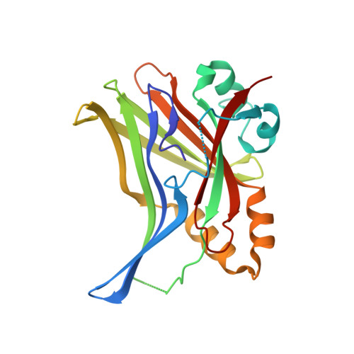
    The dysregulated Hippo pathway and, consequently, hyperactivity of the transcriptional YAP/TAZ-TEAD complexes is associated with diseases such as cancer. Prevention of YAP/TAZ-TEAD triggered gene transcription is an attractive strategy for therapeutic intervention. The deeply buried and conserved lipidation pocket (P-site) of the TEAD transcription factors is druggable. The discovery and optimization of a P-site binding fragment ( 1 ) are described. Utilizing structure-based design, enhancement in target potency was engineered into the hit, capitalizing on the established X-ray structure of TEAD1. The efforts culminated in the optimized in vivo tool MSC-4106 , which exhibited desirable potency, mouse pharmacokinetic properties, and in vivo efficacy. In close correlation to compound exposure, the time- and dose-dependent downregulation of a proximal biomarker could be shown.


  • Organizational Affiliation

    Merck Healthcare KGaA, Frankfurter Str. 250, 64293 Darmstadt, Germany.


Macromolecules
Find similar proteins by:  (by identity cutoff)  |  3D Structure
Entity ID: 1
MoleculeChains Sequence LengthOrganismDetailsImage
Transcriptional enhancer factor TEF-1
A, B
220Homo sapiensMutation(s): 0 
Gene Names: TEAD1TCF13TEF1
UniProt & NIH Common Fund Data Resources
Find proteins for P28347 (Homo sapiens)
Explore P28347 
Go to UniProtKB:  P28347
PHAROS:  P28347
GTEx:  ENSG00000187079 
Entity Groups  
Sequence Clusters30% Identity50% Identity70% Identity90% Identity95% Identity100% Identity
UniProt GroupP28347
Sequence Annotations
Expand
  • Reference Sequence
Experimental Data & Validation

Experimental Data

  • Method: X-RAY DIFFRACTION
  • Resolution: 2.19 Å
  • R-Value Free:  0.236 (Depositor), 0.230 (DCC) 
  • R-Value Work:  0.190 (Depositor), 0.190 (DCC) 
  • R-Value Observed: 0.193 (Depositor) 
Space Group: P 41 21 2
Unit Cell:
Length ( Å )Angle ( ˚ )
a = 108.359α = 90
b = 108.359β = 90
c = 132.162γ = 90
Software Package:
Software NamePurpose
BUSTERrefinement
PDB_EXTRACTdata extraction
XDSdata reduction
autoPROCdata scaling
PHASERphasing

Structure Validation

View Full Validation Report


Ligand Structure Quality Assessment 

Created with Raphaël 2.3.0Worse 01 BetterLigand structure goodness of fit to experimental dataBest fitted JJUClick on this verticalbar to view details

Entry History & Funding Information

Revision History  (Full details and data files)

  • Version 1.0: 2022-07-13
    Type: Initial release
  • Version 1.1: 2022-07-27
    Changes: Database references
  • Version 1.2: 2024-01-31
    Changes: Data collection, Refinement description