7XPR | pdb_00007xpr

Crystal structrue of SeMet-MtdL:GDP


Experimental Data Snapshot

  • Method: X-RAY DIFFRACTION
  • Resolution: 2.10 Å
  • R-Value Free: 
    0.243 (Depositor), 0.242 (DCC) 
  • R-Value Work: 
    0.211 (Depositor), 0.211 (DCC) 
  • R-Value Observed: 
    0.212 (Depositor) 

wwPDB Validation 3D Report Full Report

Validation slider image for 7XPR

Ligand Structure Quality Assessment 


This is version 1.1 of the entry. See complete history

Literature

Structures of the NDP-pyranose mutase belonging to glycosyltransferase family 75 reveal residues important for Mn 2+ coordination and substrate binding.

Du, X.Chu, X.Liu, N.Jia, X.Peng, H.Xiao, Y.Liu, L.Yu, H.Li, F.He, C.

(2023) J Biological Chem 299: 102903-102903

  • DOI: https://doi.org/10.1016/j.jbc.2023.102903
  • Primary Citation Related Structures: 
    7XPR, 7XPS, 7XPT, 7XPU, 7XPV, 8HL8

  • PubMed Abstract: 

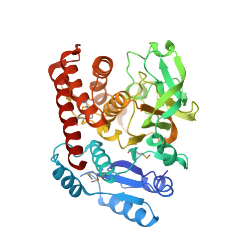
    Members of glycosyltransferase family 75 (GT75) not only reversibly catalyze the autoglycosylation of a conserved arginine residue with specific NDP-sugars but also exhibit NDP-pyranose mutase activity that reversibly converts specific NDP-pyranose to NDP-furanose. The latter activity provides valuable NDP-furanosyl donors for glycosyltransferases and requires a divalent cation as a cofactor instead of FAD used by UDP-D-galactopyranose mutase. However, details of the mechanism for NDP-pyranose mutase activity are not clear. Here we report the first crystal structures of GT75 family NDP-pyranose mutases. The novel structures of GT75 member MtdL in complex with Mn 2+ and GDP, GDP-D-glucopyranose, GDP-L-fucopyranose, GDP-L-fucofuranose, respectively, combined with site-directed mutagenesis studies, reveal key residues involved in Mn 2+ coordination, substrate binding, and catalytic reactions. We also provide a possible catalytic mechanism for this unique type of NDP-pyranose mutase. Taken together, our results highlight key elements of an enzyme family important for furanose biosynthesis.


  • Organizational Affiliation
    • Anhui Key Laboratory of Modern Biomanufacturing and School of Life Sciences, Anhui University, Hefei, Anhui, China.

Macromolecule Content 

  • Total Structure Weight: 85.13 kDa 
  • Atom Count: 6,252 
  • Modeled Residue Count: 742 
  • Deposited Residue Count: 754 
  • Unique protein chains: 1

Macromolecules

Find similar proteins by:|  3D Structure
Entity ID: 1
MoleculeChains  Sequence LengthOrganismDetailsImage
Transglycosylse
A, B
377Marinactinospora thermotoleransMutation(s): 0 
UniProt
Find proteins for G8HX37 (Marinactinospora thermotolerans)
Explore G8HX37 
Go to UniProtKB:  G8HX37
Entity Groups
Sequence Clusters30% Identity50% Identity70% Identity90% Identity95% Identity100% Identity
UniProt GroupG8HX37
Sequence Annotations
Expand
Reference Sequence

Small Molecules

Modified Residues  1 Unique
IDChains TypeFormula2D DiagramParent
MSE
Query on MSE
A, B
L-PEPTIDE LINKINGC5 H11 N O2 SeMET

Experimental Data & Validation

Experimental Data

  • Method: X-RAY DIFFRACTION
  • Resolution: 2.10 Å
  • R-Value Free:  0.243 (Depositor), 0.242 (DCC) 
  • R-Value Work:  0.211 (Depositor), 0.211 (DCC) 
  • R-Value Observed: 0.212 (Depositor) 
Space Group: C 1 2 1
Unit Cell:
Length ( Å )Angle ( ˚ )
a = 162.213α = 90
b = 97.483β = 97.32
c = 57.923γ = 90
Software Package:
Software NamePurpose
PHENIXrefinement
SCALAdata scaling
PDB_EXTRACTdata extraction
XDSdata reduction
SHELXCDphasing

Structure Validation

View Full Validation Report


Ligand Structure Quality Assessment 


Entry History 

& Funding Information

Deposition Data


Funding OrganizationLocationGrant Number
National Natural Science Foundation of China (NSFC)China--

Revision History  (Full details and data files)

  • Version 1.0: 2023-03-29
    Type: Initial release
  • Version 1.1: 2024-11-06
    Changes: Data collection, Structure summary