7XN6 | pdb_00007xn6

Cryo-EM structure of CopC-CaM-caspase-3 with ADPR-deacylization


Experimental Data Snapshot

  • Method: ELECTRON MICROSCOPY
  • Resolution: 3.45 Å
  • Aggregation State: PARTICLE 
  • Reconstruction Method: SINGLE PARTICLE 

wwPDB Validation 3D Report Full Report

Validation slider image for 7XN6

This is version 2.1 of the entry. See complete history

Literature

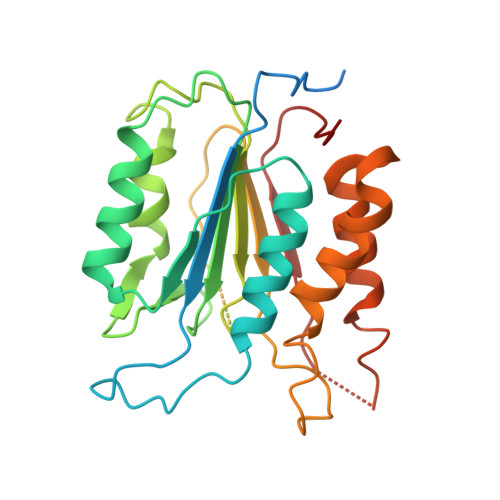
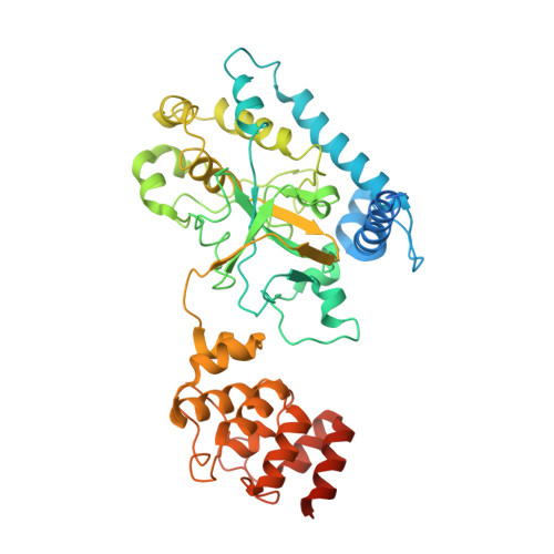
Structural insights into caspase ADPR deacylization catalyzed by a bacterial effector and host calmodulin.

Zhang, K.Peng, T.Tao, X.Tian, M.Li, Y.Wang, Z.Ma, S.Hu, S.Pan, X.Xue, J.Luo, J.Wu, Q.Fu, Y.Li, S.

(2022) Mol Cell 82: 4712-4726.e7

  • DOI: https://doi.org/10.1016/j.molcel.2022.10.032
  • Primary Citation Related Structures: 
    7XN4, 7XN5, 7XN6

  • PubMed Abstract: 

    Programmed cell death and caspase proteins play a pivotal role in host innate immune response combating pathogen infections. Blocking cell death is employed by many bacterial pathogens as a universal virulence strategy. CopC family type III effectors, including CopC from an environmental pathogen Chromobacterium violaceum, utilize calmodulin (CaM) as a co-factor to inactivate caspases by arginine ADPR deacylization. However, the molecular basis of the catalytic and substrate/co-factor binding mechanism is unknown. Here, we determine successive cryo-EM structures of CaM-CopC-caspase-3 ternary complex in pre-reaction, transition, and post-reaction states, which elucidate a multistep enzymatic mechanism of CopC-catalyzed ADPR deacylization. Moreover, we capture a snapshot of the detachment of modified caspase-3 from CopC. These structural insights are validated by mutagenesis analyses of CopC-mediated ADPR deacylization in vitro and animal infection in vivo. Our study offers a structural framework for understanding the molecular basis of arginine ADPR deacylization catalyzed by the CopC family.


  • Organizational Affiliation
    • School of Medicine, Southern University of Science and Technology, Shenzhen 518055, Guangdong, China; Institute of Infection and Immunity, Taihe Hospital, Hubei University of Medicine, Shiyan 442000, Hubei, China.

Macromolecule Content 

  • Total Structure Weight: 134.31 kDa 
  • Atom Count: 6,874 
  • Modeled Residue Count: 861 
  • Deposited Residue Count: 1,190 
  • Unique protein chains: 3

Macromolecules

Find similar proteins by:|  3D Structure
Entity ID: 1
MoleculeChains  Sequence LengthOrganismDetailsImage
Caspase-3
A, C
277Homo sapiensMutation(s): 0 
Gene Names: CASP3CPP32
EC: 3.4.22.56
UniProt & NIH Common Fund Data Resources
Find proteins for P42574 (Homo sapiens)
Explore P42574 
Go to UniProtKB:  P42574
PHAROS:  P42574
GTEx:  ENSG00000164305 
Entity Groups
Sequence Clusters30% Identity50% Identity70% Identity90% Identity95% Identity100% Identity
UniProt GroupP42574
Sequence Annotations
Expand
Reference Sequence
Find similar proteins by:|  3D Structure
Entity ID: 2
MoleculeChains  Sequence LengthOrganismDetailsImage
Arginine ADP-riboxanase CopC487Chromobacterium violaceumMutation(s): 0 
Gene Names: copCCV_2038
EC: 4.3.99
UniProt
Find proteins for Q7NWF2 (Chromobacterium violaceum (strain ATCC 12472 / DSM 30191 / JCM 1249 / CCUG 213 / NBRC 12614 / NCIMB 9131 / NCTC 9757 / MK))
Explore Q7NWF2 
Go to UniProtKB:  Q7NWF2
Entity Groups
Sequence Clusters30% Identity50% Identity70% Identity90% Identity95% Identity100% Identity
UniProt GroupQ7NWF2
Sequence Annotations
Expand
Reference Sequence
Find similar proteins by:|  3D Structure
Entity ID: 3
MoleculeChains  Sequence LengthOrganismDetailsImage
Calmodulin-1149Homo sapiensMutation(s): 0 
Gene Names: CALM1CALMCAMCAM1
UniProt & NIH Common Fund Data Resources
Find proteins for P0DP23 (Homo sapiens)
Explore P0DP23 
Go to UniProtKB:  P0DP23
PHAROS:  P0DP23
GTEx:  ENSG00000198668 
Entity Groups
Sequence Clusters30% Identity50% Identity70% Identity90% Identity95% Identity100% Identity
UniProt GroupP0DP23
Sequence Annotations
Expand
Reference Sequence

Small Molecules

Ligands 1 Unique
IDChains Name / Formula / InChI Key2D Diagram3D Interactions
NCA

Query on NCA


Download:Ideal Coordinates CCD File
E [auth B]NICOTINAMIDE
C6 H6 N2 O
DFPAKSUCGFBDDF-UHFFFAOYSA-N
Modified Residues  1 Unique
IDChains TypeFormula2D DiagramParent
A1LTQ
Query on A1LTQ
A, C
L-PEPTIDE LINKINGC21 H32 N8 O15 P2ARG

Experimental Data & Validation

Experimental Data

  • Method: ELECTRON MICROSCOPY
  • Resolution: 3.45 Å
  • Aggregation State: PARTICLE 
  • Reconstruction Method: SINGLE PARTICLE 

Structure Validation

View Full Validation Report


Entry History 

& Funding Information

Deposition Data


Funding OrganizationLocationGrant Number
Not funded--

Revision History  (Full details and data files)

  • Version 1.0: 2022-12-14
    Type: Initial release
  • Version 1.1: 2022-12-28
    Changes: Database references
  • Version 2.0: 2024-07-03
    Changes: Advisory, Atomic model, Data collection, Derived calculations, Non-polymer description, Other, Polymer sequence, Structure summary
  • Version 2.1: 2024-10-30
    Changes: Data collection, Structure summary