7X6R | pdb_00007x6r

Crystal structure of actinomycin D-echinomycin-d(AGCACGT/ACGGGCT) complex


Experimental Data Snapshot

  • Method: X-RAY DIFFRACTION
  • Resolution: 1.89 Å
  • R-Value Free: 
    0.259 (Depositor), 0.262 (DCC) 
  • R-Value Work: 
    0.244 (Depositor), 0.245 (DCC) 
  • R-Value Observed: 
    0.246 (Depositor) 

Starting Model: experimental
View more details

wwPDB Validation 3D Report Full Report

Validation slider image for 7X6R

Ligand Structure Quality Assessment 


This is version 3.0 of the entry. See complete history

Literature

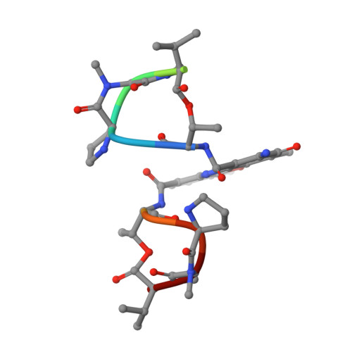
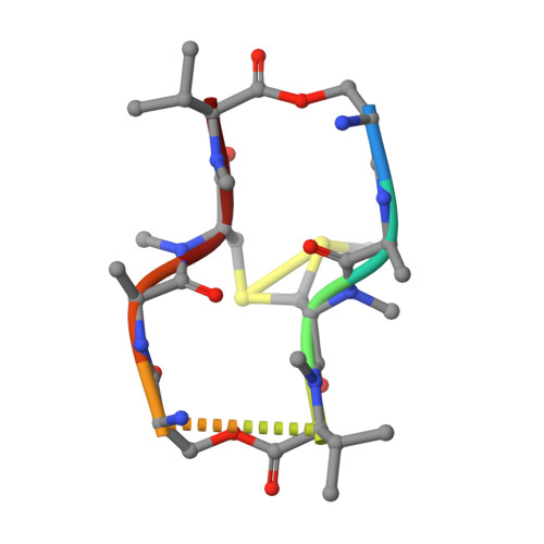
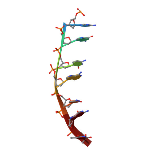
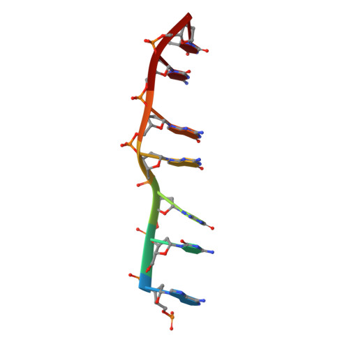
Staggered intercalation of DNA duplexes with base-pair modulation by two distinct drug molecules induces asymmetric backbone twisting and structure polymorphism.

Satange, R.Kao, S.H.Chien, C.M.Chou, S.H.Lin, C.C.Neidle, S.Hou, M.H.

(2022) Nucleic Acids Res 50: 8867-8881

  • DOI: https://doi.org/10.1093/nar/gkac629
  • Primary Citation Related Structures: 
    7X6R, 7X97, 7X9F, 7XDJ

  • PubMed Abstract: 

    The use of multiple drugs simultaneously targeting DNA is a promising strategy in cancer therapy for potentially overcoming single drug resistance. In support of this concept, we report that a combination of actinomycin D (ActD) and echinomycin (Echi), can interact in novel ways with native and mismatched DNA sequences, distinct from the structural effects produced by either drug alone. Changes in the former with GpC and CpG steps separated by a A:G or G:A mismatch or in a native DNA with canonical G:C and C:G base pairs, result in significant asymmetric backbone twists through staggered intercalation and base pair modulations. A wobble or Watson-Crick base pair at the two drug-binding interfaces can result in a single-stranded 'chair-shaped' DNA duplex with a straight helical axis. However, a novel sugar-edged hydrogen bonding geometry in the G:A mismatch leads to a 'curved-shaped' duplex. Two non-canonical G:C Hoogsteen base pairings produce a sharply kinked duplex in different forms and a four-way junction-like superstructure, respectively. Therefore, single base pair modulations on the two drug-binding interfaces could significantly affect global DNA structure. These structures thus provide a rationale for atypical DNA recognition via multiple DNA intercalators and a structural basis for the drugs' potential synergetic use.


  • Organizational Affiliation
    • Institute of Genomics and Bioinformatics, National Chung Hsing University, Taichung 402, Taiwan.

Macromolecule Content 

  • Total Structure Weight: 6.89 kDa 
  • Atom Count: 482 
  • Modeled Residue Count: 33 
  • Deposited Residue Count: 33 
  • Unique protein chains: 2
  • Unique nucleic acid chains: 2

Macromolecules


Find similar proteins by:  Sequence   |   3D Structure  
Entity ID: 3
MoleculeChains  Sequence LengthOrganismDetailsImage
Actinomycin D11StreptomycesMutation(s): 0 
Find similar proteins by:  Sequence   |   3D Structure  
Entity ID: 4
MoleculeChains  Sequence LengthOrganismDetailsImage
DSN-ALA-N2C-MVA-DSN-ALA-NCY-MVA8StreptomycesMutation(s): 0 
Find similar nucleic acids by:  Sequence
Entity ID: 1
MoleculeChains LengthOrganismImage
DNA (5'-D(P*AP*GP*CP*AP*CP*GP*T)-3')7synthetic construct
Sequence Annotations
Expand
Reference Sequence
Find similar nucleic acids by:  Sequence
Entity ID: 2
MoleculeChains LengthOrganismImage
DNA (5'-D(P*AP*CP*GP*GP*GP*CP*T)-3')7synthetic construct
Sequence Annotations
Expand
Reference Sequence

Biologically Interesting Molecules (External Reference) 

2 Unique

Experimental Data & Validation

Experimental Data

  • Method: X-RAY DIFFRACTION
  • Resolution: 1.89 Å
  • R-Value Free:  0.259 (Depositor), 0.262 (DCC) 
  • R-Value Work:  0.244 (Depositor), 0.245 (DCC) 
  • R-Value Observed: 0.246 (Depositor) 
Space Group: P 4 2 2
Unit Cell:
Length ( Å )Angle ( ˚ )
a = 43.225α = 90
b = 43.225β = 90
c = 65.839γ = 90
Software Package:
Software NamePurpose
HKL-2000data scaling
PHENIXrefinement
PDB_EXTRACTdata extraction
HKL-2000data reduction
PHENIXphasing

Structure Validation

View Full Validation Report


Ligand Structure Quality Assessment 


Entry History 

& Funding Information

Deposition Data


Funding OrganizationLocationGrant Number
Ministry of Science and Technology (MoST, Taiwan)Taiwan109-2628-M-005-001-MY4
Ministry of Science and Technology (MoST, Taiwan)Taiwan109-2311-B-005-007-MY3

Revision History  (Full details and data files)

  • Version 1.0: 2022-12-14
    Type: Initial release
  • Version 2.0: 2023-11-15
    Changes: Atomic model, Data collection, Derived calculations
  • Version 2.1: 2023-11-29
    Changes: Refinement description
  • Version 3.0: 2024-07-10
    Changes: Data collection, Derived calculations, Non-polymer description, Structure summary