7WZX | pdb_00007wzx

The structure of a Twitch Radical SAM Dehydrogenase SpeY


Experimental Data Snapshot

  • Method: X-RAY DIFFRACTION
  • Resolution: 1.98 Å
  • R-Value Free: 
    0.234 (Depositor), 0.238 (DCC) 
  • R-Value Work: 
    0.209 (Depositor), 0.213 (DCC) 
  • R-Value Observed: 
    0.210 (Depositor) 

Starting Model: experimental
View more details

wwPDB Validation 3D Report Full Report

Validation slider image for 7WZX

Ligand Structure Quality Assessment 


This is version 1.1 of the entry. See complete history

Literature

Dioxane Bridge Formation during the Biosynthesis of Spectinomycin Involves a Twitch Radical S -Adenosyl Methionine Dehydrogenase That May Have Evolved from an Epimerase.

Zhang, J.Hou, X.Chen, Z.Ko, Y.Ruszczycky, M.W.Chen, Y.Zhou, J.Liu, H.W.

(2022) J Am Chem Soc 144: 9910-9919

  • DOI: https://doi.org/10.1021/jacs.2c02676
  • Primary Citation Related Structures: 
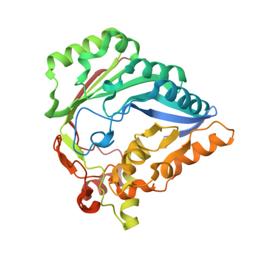
    7WZV, 7WZX, 7X0B

  • PubMed Abstract: 

    Spectinomycin is a dioxane-bridged, tricyclic aminoglycoside produced by Streptomyces spectabilis ATCC 27741. While the spe biosynthetic gene cluster for spectinomycin has been reported, the chemistry underlying construction of the dioxane ring is unknown. The twitch radical SAM enzyme SpeY from the spe cluster is shown here to catalyze dehydrogenation of the C2' alcohol of (2' R ,3' S )-tetrahydrospectinomycin to yield (3' S )-dihydrospectinomycin as a likely biosynthetic intermediate. This reaction is radical-mediated and initiated via H atom abstraction from C2' of the substrate by the 5'-deoxyadenosyl radical equivalent generated upon reductive cleavage of SAM. Crystallographic analysis of the ternary Michaelis complex places serine-183 adjacent to C2' of the bound substrate opposite C5' of SAM. Mutation of this residue to cysteine converts SpeY to the corresponding C2' epimerase mirroring the opposite phenomenon observed in the homologous twitch radical SAM epimerase HygY from the hygromycin B biosynthetic pathway. Phylogenetic analysis suggests a relatively recent evolutionary branching of putative twitch radical SAM epimerases bearing homologous cysteine residues to generate the SpeY clade of enzymes.


  • Organizational Affiliation
    • Department of Chemistry, University of Texas at Austin, Austin, Texas 78712, United States.

Macromolecule Content 

  • Total Structure Weight: 38.01 kDa 
  • Atom Count: 2,544 
  • Modeled Residue Count: 300 
  • Deposited Residue Count: 322 
  • Unique protein chains: 1

Macromolecules

Find similar proteins by:|  3D Structure
Entity ID: 1
MoleculeChains  Sequence LengthOrganismDetailsImage
4Fe-4S cluster-binding domain-containing protein322Streptomyces spectabilisMutation(s): 0 
Gene Names: speYFH965_34950
UniProt
Find proteins for A8WEZ7 (Streptomyces spectabilis)
Explore A8WEZ7 
Go to UniProtKB:  A8WEZ7
Entity Groups
Sequence Clusters30% Identity50% Identity70% Identity90% Identity95% Identity100% Identity
UniProt GroupA8WEZ7
Sequence Annotations
Expand
Reference Sequence

Small Molecules

Ligands 5 Unique
IDChains Name / Formula / InChI Key2D Diagram3D Interactions
SAM

Query on SAM


Download:Ideal Coordinates CCD File
D [auth A]S-ADENOSYLMETHIONINE
C15 H22 N6 O5 S
MEFKEPWMEQBLKI-FCKMPRQPSA-N
SF4

Query on SF4


Download:Ideal Coordinates CCD File
B [auth A],
C [auth A]
IRON/SULFUR CLUSTER
Fe4 S4
LJBDFODJNLIPKO-UHFFFAOYSA-N
7PK
(Subject of Investigation/LOI)

Query on 7PK


Download:Ideal Coordinates CCD File
I [auth A](2~{S},4~{S},6~{R})-2-[(2~{S},3~{R},5~{S},6~{R})-3,5-bis(methylamino)-2,4,6-tris(oxidanyl)cyclohexyl]oxy-6-methyl-4-oxidanyl-oxan-3-one
C14 H26 N2 O7
VKGOUJWNNSMSLM-MWLBSAGXSA-N
SO4

Query on SO4


Download:Ideal Coordinates CCD File
J [auth A],
K [auth A]
SULFATE ION
O4 S
QAOWNCQODCNURD-UHFFFAOYSA-L
GOL

Query on GOL


Download:Ideal Coordinates CCD File
E [auth A],
F [auth A],
G [auth A],
H [auth A]
GLYCEROL
C3 H8 O3
PEDCQBHIVMGVHV-UHFFFAOYSA-N

Experimental Data & Validation

Experimental Data

  • Method: X-RAY DIFFRACTION
  • Resolution: 1.98 Å
  • R-Value Free:  0.234 (Depositor), 0.238 (DCC) 
  • R-Value Work:  0.209 (Depositor), 0.213 (DCC) 
  • R-Value Observed: 0.210 (Depositor) 
Space Group: P 65 2 2
Unit Cell:
Length ( Å )Angle ( ˚ )
a = 93.93α = 90
b = 93.93β = 90
c = 143.42γ = 120
Software Package:
Software NamePurpose
PHENIXrefinement
XDSdata reduction
Aimlessdata scaling
PHASERphasing

Structure Validation

View Full Validation Report


Ligand Structure Quality Assessment 


Entry History 

& Funding Information

Deposition Data


Funding OrganizationLocationGrant Number
Not funded--

Revision History  (Full details and data files)

  • Version 1.0: 2022-12-28
    Type: Initial release
  • Version 1.1: 2023-11-29
    Changes: Data collection, Refinement description