7VVM

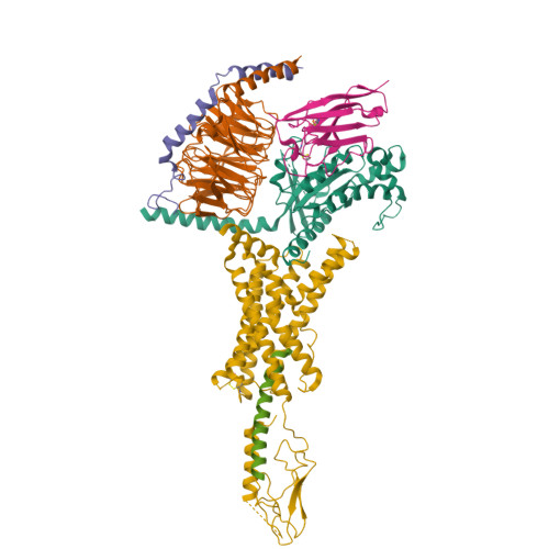
PTH-bound human PTH1R in complex with Gs (class3)


Experimental Data Snapshot

  • Method: ELECTRON MICROSCOPY
  • Resolution: 3.20 Å
  • Aggregation State: PARTICLE 
  • Reconstruction Method: SINGLE PARTICLE 

wwPDB Validation   3D Report Full Report


This is version 1.2 of the entry. See complete history


Literature

Endogenous ligand recognition and structural transition of a human PTH receptor.

Kobayashi, K.Kawakami, K.Kusakizako, T.Miyauchi, H.Tomita, A.Kobayashi, K.Shihoya, W.Yamashita, K.Nishizawa, T.Kato, H.E.Inoue, A.Nureki, O.

(2022) Mol Cell 82: 3468-3483.e5

  • DOI: https://doi.org/10.1016/j.molcel.2022.07.003
  • Primary Citation of Related Structures:  
    7VVJ, 7VVK, 7VVL, 7VVM, 7VVN, 7VVO

  • PubMed Abstract: 

    Endogenous parathyroid hormone (PTH) and PTH-related peptide (PTHrP) bind to the parathyroid hormone receptor 1 (PTH1R) and activate the stimulatory G-protein (Gs) signaling pathway. Intriguingly, the two ligands have distinct signaling and physiological properties: PTH evokes prolonged Gs activation, whereas PTHrP evokes transient Gs activation with reduced bone-resorption effects. The distinct molecular actions are ascribed to the differences in ligand recognition and dissociation kinetics. Here, we report cryoelectron microscopic structures of six forms of the human PTH1R-Gs complex in the presence of PTH or PTHrP at resolutions of 2.8 -4.1 Å. A comparison of the PTH-bound and PTHrP-bound structures reveals distinct ligand-receptor interactions underlying the ligand affinity and selectivity. Furthermore, five distinct PTH-bound structures, combined with computational analyses, provide insights into the unique and complex process of ligand dissociation from the receptor and shed light on the distinct durations of signaling induced by PTH and PTHrP.


  • Organizational Affiliation

    Department of Biological Sciences, Graduate School of Science, the University of Tokyo, Bunkyo, Tokyo 113-0033, Japan.


Macromolecules
Find similar proteins by:  (by identity cutoff)  |  3D Structure
Entity ID: 1
MoleculeChains Sequence LengthOrganismDetailsImage
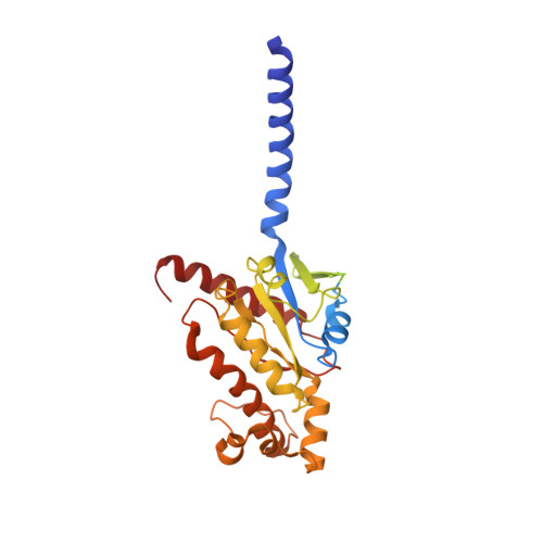
Guanine nucleotide-binding protein G(s) subunit alpha isoforms short380Homo sapiensMutation(s): 7 
Gene Names: GNASGNAS1GSP
EC: 3.6.5
Membrane Entity: Yes 
UniProt & NIH Common Fund Data Resources
Find proteins for P63092 (Homo sapiens)
Explore P63092 
Go to UniProtKB:  P63092
PHAROS:  P63092
GTEx:  ENSG00000087460 
Entity Groups  
Sequence Clusters30% Identity50% Identity70% Identity90% Identity95% Identity100% Identity
UniProt GroupP63092
Sequence Annotations
Expand
  • Reference Sequence
Find similar proteins by:  (by identity cutoff)  |  3D Structure
Entity ID: 2
MoleculeChains Sequence LengthOrganismDetailsImage
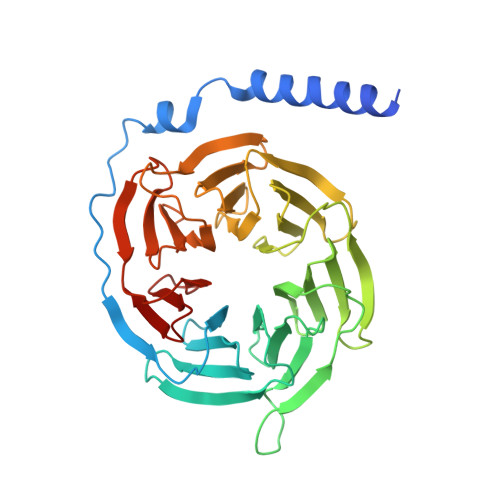
Guanine nucleotide-binding protein G(I)/G(S)/G(T) subunit beta-1351Rattus norvegicusMutation(s): 0 
Gene Names: Gnb1
UniProt
Find proteins for P54311 (Rattus norvegicus)
Explore P54311 
Go to UniProtKB:  P54311
Entity Groups  
Sequence Clusters30% Identity50% Identity70% Identity90% Identity95% Identity100% Identity
UniProt GroupP54311
Sequence Annotations
Expand
  • Reference Sequence
Find similar proteins by:  (by identity cutoff)  |  3D Structure
Entity ID: 3
MoleculeChains Sequence LengthOrganismDetailsImage
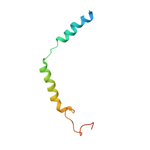
Guanine nucleotide-binding protein G(I)/G(S)/G(O) subunit gamma-2C [auth G]68Bos taurusMutation(s): 0 
Gene Names: GNG2
Membrane Entity: Yes 
UniProt
Find proteins for P63212 (Bos taurus)
Explore P63212 
Go to UniProtKB:  P63212
Entity Groups  
Sequence Clusters30% Identity50% Identity70% Identity90% Identity95% Identity100% Identity
UniProt GroupP63212
Sequence Annotations
Expand
  • Reference Sequence
Find similar proteins by:  (by identity cutoff)  |  3D Structure
Entity ID: 4
MoleculeChains Sequence LengthOrganismDetailsImage
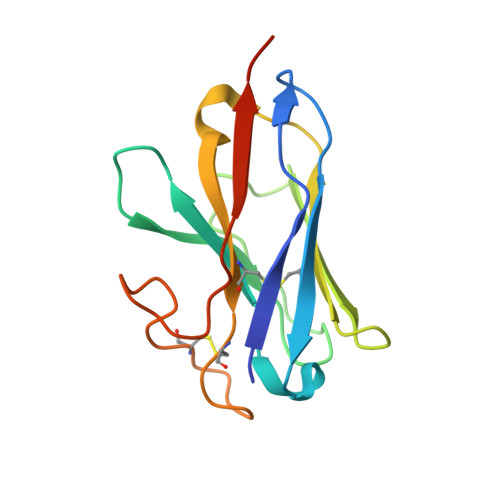
nanobody Nb35D [auth N]137unidentifiedMutation(s): 0 
Entity Groups  
Sequence Clusters30% Identity50% Identity70% Identity90% Identity95% Identity100% Identity
Sequence Annotations
Expand
  • Reference Sequence
Find similar proteins by:  (by identity cutoff)  |  3D Structure
Entity ID: 5
MoleculeChains Sequence LengthOrganismDetailsImage
Parathyroid hormoneE [auth P]34Homo sapiensMutation(s): 0 
Membrane Entity: Yes 
UniProt & NIH Common Fund Data Resources
Find proteins for P01270 (Homo sapiens)
Explore P01270 
Go to UniProtKB:  P01270
PHAROS:  P01270
GTEx:  ENSG00000152266 
Entity Groups  
Sequence Clusters30% Identity50% Identity70% Identity90% Identity95% Identity100% Identity
UniProt GroupP01270
Sequence Annotations
Expand
  • Reference Sequence
Find similar proteins by:  (by identity cutoff)  |  3D Structure
Entity ID: 6
MoleculeChains Sequence LengthOrganismDetailsImage
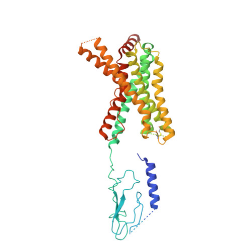
Parathyroid hormone/parathyroid hormone-related peptide receptorF [auth R]473Homo sapiensMutation(s): 0 
Gene Names: PTH1RPTHRPTHR1
Membrane Entity: Yes 
UniProt & NIH Common Fund Data Resources
Find proteins for Q03431 (Homo sapiens)
Explore Q03431 
Go to UniProtKB:  Q03431
PHAROS:  Q03431
GTEx:  ENSG00000160801 
Entity Groups  
Sequence Clusters30% Identity50% Identity70% Identity90% Identity95% Identity100% Identity
UniProt GroupQ03431
Sequence Annotations
Expand
  • Reference Sequence
Experimental Data & Validation

Experimental Data

  • Method: ELECTRON MICROSCOPY
  • Resolution: 3.20 Å
  • Aggregation State: PARTICLE 
  • Reconstruction Method: SINGLE PARTICLE 

Structure Validation

View Full Validation Report


Entry History & Funding Information

Deposition Data


Funding OrganizationLocationGrant Number
Japan Society for the Promotion of Science (JSPS)Japan--

Revision History  (Full details and data files)

  • Version 1.0: 2022-08-03
    Type: Initial release
  • Version 1.1: 2023-02-15
    Changes: Database references
  • Version 1.2: 2024-10-30
    Changes: Data collection, Structure summary