7VGU

Time-resolved serial femtosecond crystallography structure of light-driven chloride ion-pumping rhodopsin, NM-R3 : structure obtained 1 msec after photoexcitation with bromide ion


Experimental Data Snapshot

  • Method: X-RAY DIFFRACTION
  • Resolution: 2.10 Å
  • R-Value Free: 0.211 
  • R-Value Work: 0.182 
  • R-Value Observed: 0.183 

Starting Model: experimental
View more details

wwPDB Validation   3D Report Full Report


Ligand Structure Quality Assessment 


This is version 1.3 of the entry. See complete history


Literature

Conformational alterations in unidirectional ion transport of a light-driven chloride pump revealed using X-ray free electron lasers.

Hosaka, T.Nomura, T.Kubo, M.Nakane, T.Fangjia, L.Sekine, S.I.Ito, T.Murayama, K.Ihara, K.Ehara, H.Kashiwagi, K.Katsura, K.Akasaka, R.Hisano, T.Tanaka, T.Tanaka, R.Arima, T.Yamashita, A.Sugahara, M.Naitow, H.Matsuura, Y.Yoshizawa, S.Tono, K.Owada, S.Nureki, O.Kimura-Someya, T.Iwata, S.Nango, E.Shirouzu, M.

(2022) Proc Natl Acad Sci U S A 119

  • DOI: https://doi.org/10.1073/pnas.2117433119
  • Primary Citation of Related Structures:  
    7VGT, 7VGU, 7VGV

  • PubMed Abstract: 

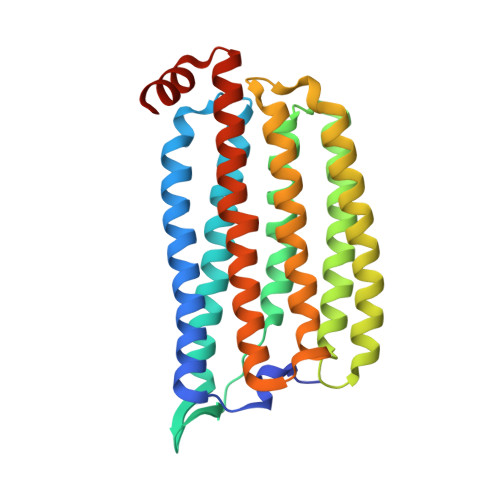
    Light-driven chloride-pumping rhodopsins actively transport anions, including various halide ions, across cell membranes. Recent studies using time-resolved serial femtosecond crystallography (TR-SFX) have uncovered the structural changes and ion transfer mechanisms in light-driven cation-pumping rhodopsins. However, the mechanism by which the conformational changes pump an anion to achieve unidirectional ion transport, from the extracellular side to the cytoplasmic side, in anion-pumping rhodopsins remains enigmatic. We have collected TR-SFX data of Nonlabens marinus rhodopsin-3 (NM-R3), derived from a marine flavobacterium, at 10-µs and 1-ms time points after photoexcitation. Our structural analysis reveals the conformational alterations during ion transfer and after ion release. Movements of the retinal chromophore initially displace a conserved tryptophan to the cytoplasmic side of NM-R3, accompanied by a slight shift of the halide ion bound to the retinal. After ion release, the inward movements of helix C and helix G and the lateral displacements of the retinal block access to the extracellular side of NM-R3. Anomalous signal data have also been obtained from NM-R3 crystals containing iodide ions. The anomalous density maps provide insight into the halide binding site for ion transfer in NM-R3.


  • Organizational Affiliation

    RIKEN Center for Biosystems Dynamics Research, Yokohama, Kanagawa 230-0045, Japan.


Macromolecules
Find similar proteins by:  (by identity cutoff)  |  3D Structure
Entity ID: 1
MoleculeChains Sequence LengthOrganismDetailsImage
Chloride pumping rhodopsin279Nonlabens marinus S1-08Mutation(s): 0 
Gene Names: ClRNMS_1267
Membrane Entity: Yes 
UniProt
Find proteins for W8VZW3 (Nonlabens marinus S1-08)
Explore W8VZW3 
Go to UniProtKB:  W8VZW3
Entity Groups  
Sequence Clusters30% Identity50% Identity70% Identity90% Identity95% Identity100% Identity
UniProt GroupW8VZW3
Sequence Annotations
Expand
  • Reference Sequence
Small Molecules
Ligands 4 Unique
IDChains Name / Formula / InChI Key2D Diagram3D Interactions
RET (Subject of Investigation/LOI)
Query on RET

Download Ideal Coordinates CCD File 
B [auth A]RETINAL
C20 H28 O
NCYCYZXNIZJOKI-OVSJKPMPSA-N
D10 (Subject of Investigation/LOI)
Query on D10

Download Ideal Coordinates CCD File 
G [auth A],
H [auth A],
I [auth A]
DECANE
C10 H22
DIOQZVSQGTUSAI-UHFFFAOYSA-N
HEX (Subject of Investigation/LOI)
Query on HEX

Download Ideal Coordinates CCD File 
C [auth A],
D [auth A],
E [auth A],
F [auth A]
HEXANE
C6 H14
VLKZOEOYAKHREP-UHFFFAOYSA-N
BR (Subject of Investigation/LOI)
Query on BR

Download Ideal Coordinates CCD File 
J [auth A],
K [auth A],
L [auth A]
BROMIDE ION
Br
CPELXLSAUQHCOX-UHFFFAOYSA-M
Experimental Data & Validation

Experimental Data

Unit Cell:
Length ( Å )Angle ( ˚ )
a = 104.3α = 90
b = 51.1β = 131.5
c = 78.4γ = 90
Software Package:
Software NamePurpose
PHENIXrefinement
PDB_EXTRACTdata extraction
CrystFELdata reduction
CrystFELdata scaling
PHENIXphasing

Structure Validation

View Full Validation Report


Ligand Structure Quality Assessment 


Entry History & Funding Information

Deposition Data


Funding OrganizationLocationGrant Number
Not funded--

Revision History  (Full details and data files)

  • Version 1.0: 2022-02-16
    Type: Initial release
  • Version 1.1: 2022-03-09
    Changes: Database references
  • Version 1.2: 2023-09-06
    Changes: Data collection, Database references
  • Version 1.3: 2023-11-29
    Changes: Refinement description