7V1V | pdb_00007v1v

Difructose dianhydride I synthase/hydrolase (alphaFFase1) from Bifidobacterium dentium, ligand-free form


Experimental Data Snapshot

  • Method: X-RAY DIFFRACTION
  • Resolution: 1.96 Å
  • R-Value Free: 
    0.191 (Depositor), 0.197 (DCC) 
  • R-Value Work: 
    0.153 (Depositor), 0.162 (DCC) 
  • R-Value Observed: 
    0.155 (Depositor) 

Starting Model: experimental
View more details

wwPDB Validation 3D Report Full Report

Validation slider image for 7V1V

This is version 1.2 of the entry. See complete history

Literature

Identification of difructose dianhydride I synthase/hydrolase from an oral bacterium establishes a novel glycoside hydrolase family.

Kashima, T.Okumura, K.Ishiwata, A.Kaieda, M.Terada, T.Arakawa, T.Yamada, C.Shimizu, K.Tanaka, K.Kitaoka, M.Ito, Y.Fujita, K.Fushinobu, S.

(2021) J Biological Chem 297: 101324-101324

  • DOI: https://doi.org/10.1016/j.jbc.2021.101324
  • Primary Citation Related Structures: 
    7V1V, 7V1W, 7V1X

  • PubMed Abstract: 

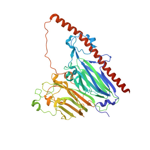
    Fructooligosaccharides and their anhydrides are widely used as health-promoting foods and prebiotics. Various enzymes acting on β-D-fructofuranosyl linkages of natural fructan polymers have been used to produce functional compounds. However, enzymes that hydrolyze and form α-D-fructofuranosyl linkages have been less studied. Here, we identified the BBDE_2040 gene product from Bifidobacterium dentium (α-D-fructofuranosidase and difructose dianhydride I synthase/hydrolase from Bifidobacterium dentium [αFFase1]) as an enzyme with α-D-fructofuranosidase and α-D-arabinofuranosidase activities and an anomer-retaining manner. αFFase1 is not homologous with any known enzymes, suggesting that it is a member of a novel glycoside hydrolase family. When caramelized fructose sugar was incubated with αFFase1, conversions of β-D-Frup-(2→1)-α-D-Fruf to α-D-Fruf-1,2':2,1'-β-D-Frup (diheterolevulosan II) and β-D-Fruf-(2→1)-α-D-Fruf (inulobiose) to α-D-Fruf-1,2':2,1'-β-D-Fruf (difructose dianhydride I [DFA I]) were observed. The reaction equilibrium between inulobiose and DFA I was biased toward the latter (1:9) to promote the intramolecular dehydrating condensation reaction. Thus, we named this enzyme DFA I synthase/hydrolase. The crystal structures of αFFase1 in complex with β-D-Fruf and β-D-Araf were determined at the resolutions of up to 1.76 Å. Modeling of a DFA I molecule in the active site and mutational analysis also identified critical residues for catalysis and substrate binding. The hexameric structure of αFFase1 revealed the connection of the catalytic pocket to a large internal cavity via a channel. Molecular dynamics analysis implied stable binding of DFA I and inulobiose to the active site with surrounding water molecules. Taken together, these results establish DFA I synthase/hydrolase as a member of a new glycoside hydrolase family (GH172).


  • Organizational Affiliation
    • Department of Biotechnology, The University of Tokyo, Tokyo, Japan.

Macromolecule Content 

  • Total Structure Weight: 319.05 kDa 
  • Atom Count: 24,197 
  • Modeled Residue Count: 2,678 
  • Deposited Residue Count: 2,760 
  • Unique protein chains: 1

Macromolecules

Find similar proteins by:|  3D Structure
Entity ID: 1
MoleculeChains  Sequence LengthOrganismDetailsImage
Difructose dianhydride I synthase/hydrolase (alphaFFase1)
A, B, C, D, E
A, B, C, D, E, F
460Bifidobacterium dentiumMutation(s): 0 
Gene Names: BDLFYP24_02130
UniProt
Find proteins for A0A6L9SN29 (Bifidobacterium dentium)
Explore A0A6L9SN29 
Go to UniProtKB:  A0A6L9SN29
Entity Groups
Sequence Clusters30% Identity50% Identity70% Identity90% Identity95% Identity100% Identity
UniProt GroupA0A6L9SN29
Sequence Annotations
Expand
Reference Sequence

Small Molecules

Ligands 3 Unique
IDChains Name / Formula / InChI Key2D Diagram3D Interactions
TAR

Query on TAR


Download:Ideal Coordinates CCD File
J [auth C],
N [auth E]
D(-)-TARTARIC ACID
C4 H6 O6
FEWJPZIEWOKRBE-LWMBPPNESA-N
MPD

Query on MPD


Download:Ideal Coordinates CCD File
I [auth B],
P [auth F]
(4S)-2-METHYL-2,4-PENTANEDIOL
C6 H14 O2
SVTBMSDMJJWYQN-YFKPBYRVSA-N
CA

Query on CA


Download:Ideal Coordinates CCD File
G [auth A]
H [auth B]
K [auth C]
L [auth C]
M [auth D]
G [auth A],
H [auth B],
K [auth C],
L [auth C],
M [auth D],
O [auth E]
CALCIUM ION
Ca
BHPQYMZQTOCNFJ-UHFFFAOYSA-N

Experimental Data & Validation

Experimental Data

  • Method: X-RAY DIFFRACTION
  • Resolution: 1.96 Å
  • R-Value Free:  0.191 (Depositor), 0.197 (DCC) 
  • R-Value Work:  0.153 (Depositor), 0.162 (DCC) 
  • R-Value Observed: 0.155 (Depositor) 
Space Group: P 1 21 1
Unit Cell:
Length ( Å )Angle ( ˚ )
a = 98.552α = 90
b = 156.97β = 109.93
c = 100.909γ = 90
Software Package:
Software NamePurpose
REFMACrefinement
PDB_EXTRACTdata extraction
XDSdata reduction
Aimlessdata scaling
MoRDaphasing

Structure Validation

View Full Validation Report


Entry History 

& Funding Information

Deposition Data


Funding OrganizationLocationGrant Number
Japan Society for the Promotion of Science (JSPS)Japan26660083
Japan Society for the Promotion of Science (JSPS)Japan24380053

Revision History  (Full details and data files)

  • Version 1.0: 2021-11-03
    Type: Initial release
  • Version 1.1: 2022-02-16
    Changes: Database references
  • Version 1.2: 2023-11-29
    Changes: Data collection, Refinement description