7T7L | pdb_00007t7l

Structure of human G9a SET-domain (EHMT2) in complex with covalent inhibitor (Compound 1)


Experimental Data Snapshot

  • Method: X-RAY DIFFRACTION
  • Resolution: 2.20 Å
  • R-Value Free: 
    0.251 (Depositor), 0.253 (DCC) 
  • R-Value Work: 
    0.204 (Depositor), 0.208 (DCC) 
  • R-Value Observed: 
    0.206 (Depositor) 

Starting Model: experimental
View more details

wwPDB Validation 3D Report Full Report

Validation slider image for 7T7L

Ligand Structure Quality Assessment 


This is version 1.5 of the entry. See complete history

Literature

Discovery of the First-in-Class G9a/GLP Covalent Inhibitors.

Park, K.S.Xiong, Y.Yim, H.Velez, J.Babault, N.Kumar, P.Liu, J.Jin, J.

(2022) J Med Chem 65: 10506-10522

  • DOI: https://doi.org/10.1021/acs.jmedchem.2c00652
  • Primary Citation Related Structures: 
    7T7L, 7T7M

  • PubMed Abstract: 

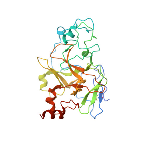
    The highly homologous protein lysine methyltransferases G9a and GLP, which catalyze mono- and dimethylation of histone H3 lysine 9 (H3K9), have been implicated in various human diseases. To investigate functions of G9a and GLP in human diseases, we and others reported several noncovalent reversible small-molecule inhibitors of G9a and GLP. Here, we report the discovery of the first-in-class G9a/GLP covalent irreversible inhibitors, 1 and 8 (MS8511), by targeting a cysteine residue at the substrate binding site. We characterized these covalent inhibitors in enzymatic, mass spectrometry based and cellular assays and using X-ray crystallography. Compared to the noncovalent G9a/GLP inhibitor UNC0642, covalent inhibitor 8 displayed improved potency in enzymatic and cellular assays. Interestingly, compound 8 also displayed potential kinetic preference for covalently modifying G9a over GLP. Collectively, compound 8 could be a useful chemical tool for studying the functional roles of G9a and GLP by covalently modifying and inhibiting these methyltransferases.


  • Organizational Affiliation
    • Mount Sinai Center for Therapeutics Discovery, Departments of Pharmacological Sciences, Oncological Sciences and Neuroscience, Tisch Cancer Institute, Icahn School of Medicine at Mount Sinai, New York, New York 10029, United States.

Macromolecule Content 

  • Total Structure Weight: 135.05 kDa 
  • Atom Count: 9,210 
  • Modeled Residue Count: 1,065 
  • Deposited Residue Count: 1,132 
  • Unique protein chains: 1

Macromolecules

Find similar proteins by:|  3D Structure
Entity ID: 1
MoleculeChains  Sequence LengthOrganismDetailsImage
Histone-lysine N-methyltransferase EHMT2
A, B, C, D
283Homo sapiensMutation(s): 0 
Gene Names: EHMT2BAT8C6orf30G9AKMT1CNG36
EC: 2.1.1 (PDB Primary Data), 2.1.1.43 (PDB Primary Data), 2.1.1.367 (UniProt)
UniProt & NIH Common Fund Data Resources
Find proteins for Q96KQ7 (Homo sapiens)
Explore Q96KQ7 
Go to UniProtKB:  Q96KQ7
PHAROS:  Q96KQ7
GTEx:  ENSG00000204371 
Entity Groups
Sequence Clusters30% Identity50% Identity70% Identity90% Identity95% Identity100% Identity
UniProt GroupQ96KQ7
Sequence Annotations
Expand
Reference Sequence

Small Molecules

Ligands 4 Unique
IDChains Name / Formula / InChI Key2D Diagram3D Interactions
G5U
(Subject of Investigation/LOI)

Query on G5U


Download:Ideal Coordinates CCD File
BA [auth D],
J [auth A],
P [auth B]
N-(6-methoxy-4-{[1-(propan-2-yl)piperidin-4-yl]amino}-7-[3-(pyrrolidin-1-yl)propoxy]quinazolin-2-yl)propanamide
C27 H42 N6 O3
GCPRIPZOIVSIDT-UHFFFAOYSA-N
G4R
(Subject of Investigation/LOI)

Query on G4R


Download:Ideal Coordinates CCD File
V [auth C]N-(6-methoxy-4-{[1-(propan-2-yl)piperidin-4-yl]amino}-7-[3-(pyrrolidin-1-yl)propoxy]quinazolin-2-yl)prop-2-enamide
C27 H40 N6 O3
JBMUWBUCVSKHLE-UHFFFAOYSA-N
SAM

Query on SAM


Download:Ideal Coordinates CCD File
AA [auth D],
I [auth A],
O [auth B],
U [auth C]
S-ADENOSYLMETHIONINE
C15 H22 N6 O5 S
MEFKEPWMEQBLKI-FCKMPRQPSA-N
ZN

Query on ZN


Download:Ideal Coordinates CCD File
E [auth A]
F [auth A]
G [auth A]
H [auth A]
K [auth B]
E [auth A],
F [auth A],
G [auth A],
H [auth A],
K [auth B],
L [auth B],
M [auth B],
N [auth B],
Q [auth C],
R [auth C],
S [auth C],
T [auth C],
W [auth D],
X [auth D],
Y [auth D],
Z [auth D]
ZINC ION
Zn
PTFCDOFLOPIGGS-UHFFFAOYSA-N
Binding Affinity Annotations 
IDSourceBinding Affinity
G4R BindingDB:  7T7L Kd: 790 (nM) from 1 assay(s)
IC50: min: 100, max: 3100 (nM) from 2 assay(s)

Experimental Data & Validation

Experimental Data

  • Method: X-RAY DIFFRACTION
  • Resolution: 2.20 Å
  • R-Value Free:  0.251 (Depositor), 0.253 (DCC) 
  • R-Value Work:  0.204 (Depositor), 0.208 (DCC) 
  • R-Value Observed: 0.206 (Depositor) 
Space Group: P 1
Unit Cell:
Length ( Å )Angle ( ˚ )
a = 56.821α = 71.15
b = 72.303β = 86.17
c = 85.029γ = 89.1
Software Package:
Software NamePurpose
PHENIXrefinement
SCALAdata scaling
PDB_EXTRACTdata extraction
MOLREPphasing
MOSFLMdata reduction

Structure Validation

View Full Validation Report


Ligand Structure Quality Assessment 


Entry History 

& Funding Information

Deposition Data


Funding OrganizationLocationGrant Number
National Institutes of Health/National Cancer Institute (NIH/NCI)United StatesR01HD088626

Revision History  (Full details and data files)

  • Version 1.0: 2022-07-06
    Type: Initial release
  • Version 1.1: 2022-07-13
    Changes: Database references
  • Version 1.2: 2022-08-24
    Changes: Database references
  • Version 1.3: 2023-10-18
    Changes: Data collection, Refinement description
  • Version 1.4: 2024-03-13
    Changes: Source and taxonomy
  • Version 1.5: 2024-10-16
    Changes: Structure summary