Ex silico engineering of cystine-dense peptides yielding a potent bispecific T cell engager.
Crook, Z.R., Girard, E.J., Sevilla, G.P., Brusniak, M.Y., Rupert, P.B., Friend, D.J., Gewe, M.M., Clarke, M., Lin, I., Ruff, R., Pakiam, F., Phi, T.D., Bandaranayake, A., Correnti, C.E., Mhyre, A.J., Nairn, N.W., Strong, R.K., Olson, J.M.(2022) Sci Transl Med 14: eabn0402-eabn0402
- PubMed: 35584229 Search on PubMedSearch on PubMed Central
- DOI: https://doi.org/10.1126/scitranslmed.abn0402
- Primary Citation Related Structures:
7SAO, 7SAP, 7SGQ, 7SJQ, 7SLT, 7SNC, 7SND - PubMed Abstract:
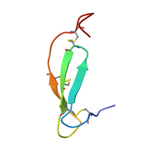
Cystine-dense peptides (CDPs) are a miniprotein class that can drug difficult targets with high affinity and low immunogenicity. Tools for their design, however, are not as developed as those for small-molecule and antibody drugs. CDPs have diverse taxonomic origins, but structural characterization is lacking. Here, we adapted Iterative Threading ASSEmbly Refinement (I-TASSER) and Rosetta protein modeling software for structural prediction of 4298 CDP scaffolds and performed in silico prescreening for CDP binders to targets of interest. Mammalian display screening of a library of docking-enriched, methionine and tyrosine scanned (DEMYS) CDPs against PD-L1 yielded binders from four distinct CDP scaffolds. One was affinity-matured, and cocrystallography yielded a high-affinity ( K D = 202 pM) PD-L1-binding CDP that competes with PD-1 for PD-L1 binding. Its subsequent incorporation into a CD3-binding bispecific T cell engager produced a molecule with pM-range in vitro T cell killing potency and which substantially extends survival in two different xenograft tumor-bearing mouse models. Both in vitro and in vivo, the CDP-incorporating bispecific molecule outperformed a comparator antibody-based molecule. This CDP modeling and DEMYS technique can accelerate CDP therapeutic development.
- Clinical Research Division, Fred Hutchinson Cancer Research Center, Seattle, WA 98109, USA.
Organizational Affiliation: