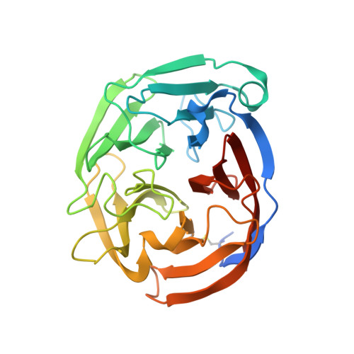
Quantitative differentiation of benign and misfolded glaucoma-causing myocilin variants on the basis of protein thermal stability.
Scelsi, H.F., Hill, K.R., Barlow, B.M., Martin, M.D., Lieberman, R.L.(2023) Dis Model Mech 16
- PubMed: 36579626
- DOI: https://doi.org/10.1242/dmm.049816
- Primary Citation of Related Structures:
7SIB, 7SIJ, 7SJT, 7SJU, 7SJV, 7SJW, 7SKD, 7SKE, 7SKF, 7SKG, 7T8D - PubMed Abstract:
Accurate predictions of the pathogenicity of mutations associated with genetic diseases are key to the success of precision medicine. Inherited missense mutations in the myocilin (MYOC) gene, within its olfactomedin (OLF) domain, constitute the strongest genetic link to primary open-angle glaucoma via a toxic gain of function, and thus MYOC is an attractive precision-medicine target. However, not all mutations in MYOC cause glaucoma, and common variants are expected to be neutral polymorphisms. The Genome Aggregation Database (gnomAD) lists ∼100 missense variants documented within OLF, all of which are relatively rare (allele frequency <0.001%) and nearly all are of unknown pathogenicity. To distinguish disease-causing OLF variants from benign OLF variants, we first characterized the most prevalent population-based variants using a suite of cellular and biophysical assays, and identified two variants with features of aggregation-prone familial disease variants. Next, we considered all available biochemical and clinical data to demonstrate that pathogenic and benign variants can be differentiated statistically based on a single metric: the thermal stability of OLF. Our results motivate genotyping MYOC in patients for clinical monitoring of this widespread, painless and irreversible ocular disease.
Organizational Affiliation:
School of Chemistry & Biochemistry, Georgia Institute of Technology, 901 Atlantic Dr. NW, Atlanta, GA 30332-0400, USA.


