7QEE | pdb_00007qee

SN243 mutant D415N bound to para-nitrophenyl-Beta-D-glucuronide


Experimental Data Snapshot

  • Method: X-RAY DIFFRACTION
  • Resolution: 2.37 Å
  • R-Value Free: 
    0.273 (Depositor), 0.270 (DCC) 
  • R-Value Work: 
    0.217 (Depositor), 0.228 (DCC) 
  • R-Value Observed: 
    0.220 (Depositor) 

Starting Model: experimental
View more details

wwPDB Validation 3D Report Full Report

Validation slider image for 7QEE

Ligand Structure Quality Assessment 


This is version 1.2 of the entry. See complete history

Literature

Functional metagenomic screening identifies an unexpected beta-glucuronidase.

Neun, S.Brear, P.Campbell, E.Tryfona, T.El Omari, K.Wagner, A.Dupree, P.Hyvonen, M.Hollfelder, F.

(2022) Nat Chem Biol 18: 1096-1103

  • DOI: https://doi.org/10.1038/s41589-022-01071-x
  • Primary Citation Related Structures: 
    7QE1, 7QE2, 7QEA, 7QEE, 7QEF, 7QG4

  • PubMed Abstract: 

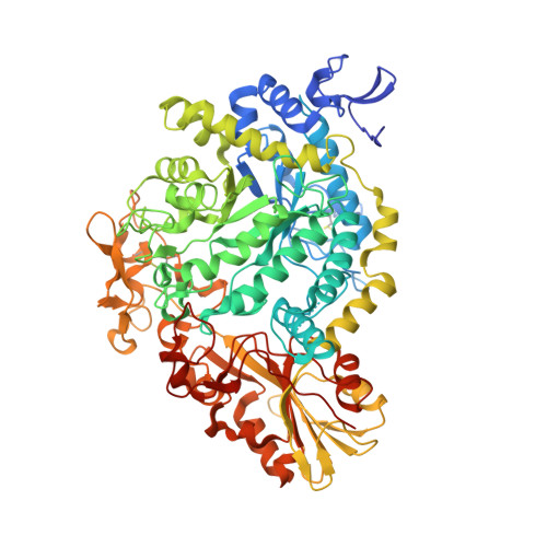
    The abundance of recorded protein sequence data stands in contrast to the small number of experimentally verified functional annotation. Here we screened a million-membered metagenomic library at ultrahigh throughput in microfluidic droplets for β-glucuronidase activity. We identified SN243, a genuine β-glucuronidase with little homology to previously studied enzymes of this type, as a glycoside hydrolase 3 family member. This glycoside hydrolase family contains only one recently added β-glucuronidase, showing that a functional metagenomic approach can shed light on assignments that are currently 'unpredictable' by bioinformatics. Kinetic analyses of SN243 characterized it as a promiscuous catalyst and structural analysis suggests regions of divergence from homologous glycoside hydrolase 3 members creating a wide-open active site. With a screening throughput of >10 7 library members per day, picolitre-volume microfluidic droplets enable functional assignments that complement current enzyme database dictionaries and provide bridgeheads for the annotation of unexplored sequence space.


  • Organizational Affiliation
    • Department of Biochemistry, University of Cambridge, Cambridge, UK.

Macromolecule Content 

  • Total Structure Weight: 166.62 kDa 
  • Atom Count: 11,451 
  • Modeled Residue Count: 1,476 
  • Deposited Residue Count: 1,518 
  • Unique protein chains: 1

Macromolecules

Find similar proteins by:|  3D Structure
Entity ID: 1
MoleculeChains  Sequence LengthOrganismDetailsImage
SN243
A, B
759synthetic constructMutation(s): 0 

Small Molecules

Ligands 4 Unique
IDChains Name / Formula / InChI Key2D Diagram3D Interactions
C3G
(Subject of Investigation/LOI)

Query on C3G


Download:Ideal Coordinates CCD File
S [auth A]4-nitrophenyl beta-D-glucopyranosiduronic acid
C12 H13 N O9
QSUILVWOWLUOEU-GOVZDWNOSA-N
BDP
(Subject of Investigation/LOI)

Query on BDP


Download:Ideal Coordinates CCD File
JA [auth B]beta-D-glucopyranuronic acid
C6 H10 O7
AEMOLEFTQBMNLQ-QIUUJYRFSA-N
SO4

Query on SO4


Download:Ideal Coordinates CCD File
HA [auth B]
IA [auth B]
O [auth A]
P [auth A]
Q [auth A]
HA [auth B],
IA [auth B],
O [auth A],
P [auth A],
Q [auth A],
R [auth A]
SULFATE ION
O4 S
QAOWNCQODCNURD-UHFFFAOYSA-L
ZN

Query on ZN


Download:Ideal Coordinates CCD File
AA [auth B]
BA [auth B]
C [auth A]
CA [auth B]
D [auth A]
AA [auth B],
BA [auth B],
C [auth A],
CA [auth B],
D [auth A],
DA [auth B],
E [auth A],
EA [auth B],
F [auth A],
FA [auth B],
G [auth A],
GA [auth B],
H [auth A],
I [auth A],
J [auth A],
K [auth A],
L [auth A],
M [auth A],
N [auth A],
T [auth B],
U [auth B],
V [auth B],
W [auth B],
X [auth B],
Y [auth B],
Z [auth B]
ZINC ION
Zn
PTFCDOFLOPIGGS-UHFFFAOYSA-N

Experimental Data & Validation

Experimental Data

  • Method: X-RAY DIFFRACTION
  • Resolution: 2.37 Å
  • R-Value Free:  0.273 (Depositor), 0.270 (DCC) 
  • R-Value Work:  0.217 (Depositor), 0.228 (DCC) 
  • R-Value Observed: 0.220 (Depositor) 
Space Group: P 1
Unit Cell:
Length ( Å )Angle ( ˚ )
a = 62.499α = 113.69
b = 82.173β = 90.65
c = 93.041γ = 90.31
Software Package:
Software NamePurpose
PHENIXrefinement
XDSdata reduction
Aimlessdata scaling
PDB_EXTRACTdata extraction
PHASERphasing

Structure Validation

View Full Validation Report


Ligand Structure Quality Assessment 


Entry History 

& Funding Information

Deposition Data


Funding OrganizationLocationGrant Number
European Union (EU)Metafluidics, 685474

Revision History  (Full details and data files)

  • Version 1.0: 2022-11-16
    Type: Initial release
  • Version 1.1: 2024-01-31
    Changes: Data collection, Refinement description
  • Version 1.2: 2024-11-20
    Changes: Structure summary