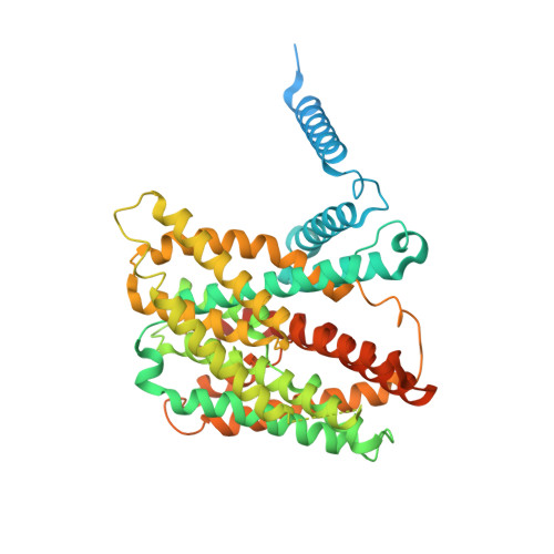
7P1J | pdb_00007p1j

Cryo EM structure of bison NHA2 in detergent structure


Experimental Data Snapshot

  • Method: ELECTRON MICROSCOPY
  • Resolution: 3.04 Å
  • Aggregation State: PARTICLE 
  • Reconstruction Method: SINGLE PARTICLE 

wwPDB Validation 3D Report Full Report

Validation slider image for 7P1J

This is version 1.4 of the entry. See complete history. 

Literature

Structure, mechanism and lipid-mediated remodeling of the mammalian Na + /H + exchanger NHA2.

Matsuoka, R., Fudim, R., Jung, S., Zhang, C., Bazzone, A., Chatzikyriakidou, Y., Robinson, C.V., Nomura, N., Iwata, S., Landreh, M., Orellana, L., Beckstein, O., Drew, D.

(2022) Nat Struct Mol Biol 29: 108-120

  • DOI: https://doi.org/10.1038/s41594-022-00738-2
  • Primary Citation Related Structures: 
    7P1I, 7P1J, 7P1K

  • PubMed Abstract: 

    The Na + /H + exchanger SLC9B2, also known as NHA2, correlates with the long-sought-after Na + /Li + exchanger linked to the pathogenesis of diabetes mellitus and essential hypertension in humans. Despite the functional importance of NHA2, structural information and the molecular basis for its ion-exchange mechanism have been lacking. Here we report the cryo-EM structures of bison NHA2 in detergent and in nanodiscs, at 3.0 and 3.5 Å resolution, respectively. The bison NHA2 structure, together with solid-state membrane-based electrophysiology, establishes the molecular basis for electroneutral ion exchange. NHA2 consists of 14 transmembrane (TM) segments, rather than the 13 TMs previously observed in mammalian Na + /H + exchangers (NHEs) and related bacterial antiporters. The additional N-terminal helix in NHA2 forms a unique homodimer interface with a large intracellular gap between the protomers, which closes in the presence of phosphoinositol lipids. We propose that the additional N-terminal helix has evolved as a lipid-mediated remodeling switch for the regulation of NHA2 activity.


  • Organizational Affiliation: 
    • Department of Biochemistry and Biophysics, Stockholm University, Stockholm, Sweden.

Macromolecule Content 

  • Total Structure Weight: 114.84 kDa 
  • Atom Count: 6,502 
  • Modeled Residue Count: 868 
  • Deposited Residue Count: 1,070 
  • Unique protein chains: 1

Macromolecules

Find similar proteins by:|  3D Structure
Entity ID: 1
MoleculeChains  Sequence LengthOrganismDetailsImage
mitochondrial sodium/hydrogen exchanger 9B2A [auth B],
B [auth A]
535Bison bisonMutation(s): 0 
Membrane Entity: Yes 
UniProt
Find proteins for A0A6P3HVI0 (Bison bison bison)
Explore A0A6P3HVI0 
Go to UniProtKB:  A0A6P3HVI0
Entity Groups
Sequence Clusters30% Identity50% Identity70% Identity90% Identity95% Identity100% Identity
UniProt GroupA0A6P3HVI0
Sequence Annotations
Expand
Reference Sequence

Experimental Data & Validation

Experimental Data

  • Method: ELECTRON MICROSCOPY
  • Resolution: 3.04 Å
  • Aggregation State: PARTICLE 
  • Reconstruction Method: SINGLE PARTICLE 
EM Software:
TaskSoftware PackageVersion
MODEL REFINEMENTPHENIX
RECONSTRUCTIONRELION3.0

Structure Validation

View Full Validation Report


Entry History 

Deposition Data

Revision History  (Full details and data files)

  • Version 1.0: 2022-01-26
    Type: Initial release
  • Version 1.1: 2022-02-02
    Changes: Structure summary
  • Version 1.2: 2022-03-02
    Changes: Database references, Structure summary
  • Version 1.3: 2024-07-17
    Changes: Data collection
  • Version 1.4: 2025-07-02
    Changes: Data collection, Structure summary