7NQA | pdb_00007nqa

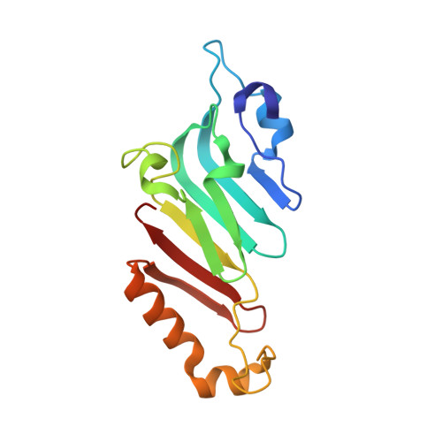
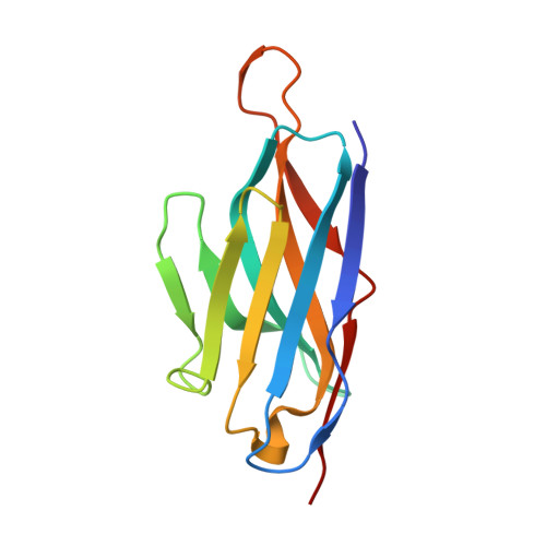
Crystal structure of Nucleoporin-98 nanobody MS98-6 complex solved at 2.2A resolution


Experimental Data Snapshot

  • Method: X-RAY DIFFRACTION
  • Resolution: 2.20 Å
  • R-Value Free: 
    0.226 (Depositor), 0.195 (DCC) 
  • R-Value Work: 
    0.176 (Depositor) 
  • R-Value Observed: 
    0.179 (Depositor) 

Starting Model: experimental
View more details

wwPDB Validation 3D Report Full Report

Validation slider image for 7NQA

Ligand Structure Quality Assessment 


This is version 1.6 of the entry. See complete history. 

Literature

A checkpoint function for Nup98 in nuclear pore formation suggested by novel inhibitory nanobodies.

Sola Colom, M., Fu, Z., Gunkel, P., Guttler, T., Trakhanov, S., Srinivasan, V., Gregor, K., Pleiner, T., Gorlich, D.

(2024) EMBO J 

  • DOI: https://doi.org/10.1038/s44318-024-00081-w
  • Primary Citation Related Structures: 
    7NOW, 7NQA, 7ZOX, 8CDS, 8CDT, 8OZB

  • PubMed Abstract: 

    Nuclear pore complex (NPC) biogenesis is a still enigmatic example of protein self-assembly. We now introduce several cross-reacting anti-Nup nanobodies for imaging intact nuclear pore complexes from frog to human. We also report a simplified assay that directly tracks postmitotic NPC assembly with added fluorophore-labeled anti-Nup nanobodies. During interphase, NPCs are inserted into a pre-existing nuclear envelope. Monitoring this process is challenging because newly assembled NPCs are indistinguishable from pre-existing ones. We overcame this problem by inserting Xenopus-derived NPCs into human nuclear envelopes and using frog-specific anti-Nup nanobodies for detection. We further asked whether anti-Nup nanobodies could serve as NPC assembly inhibitors. Using a selection strategy against conserved epitopes, we obtained anti-Nup93, Nup98, and Nup155 nanobodies that block Nup-Nup interfaces and arrest NPC assembly. We solved structures of nanobody-target complexes and identified roles for the Nup93 α-solenoid domain in recruiting Nup358 and the Nup214·88·62 complex, as well as for Nup155 and the Nup98 autoproteolytic domain in NPC scaffold assembly. The latter suggests a checkpoint linking pore formation to the assembly of the Nup98-dominated permeability barrier.


  • Organizational Affiliation: 
    • Department of Cellular Logistics, Max Planck Institute for Multidisciplinary Sciences, Göttingen, Germany.

Macromolecule Content 

  • Total Structure Weight: 61.68 kDa 
  • Atom Count: 4,657 
  • Modeled Residue Count: 552 
  • Deposited Residue Count: 552 
  • Unique protein chains: 2

Macromolecules

Find similar proteins by:|  3D Structure
Entity ID: 1
MoleculeChains  Sequence LengthOrganismDetailsImage
Nuclear pore complex protein Nup98-Nup96A,
C [auth B]
152Xenopus tropicalisMutation(s): 0 
Gene Names: nup98
UniProt
Find proteins for A0A6I8SCP2 (Xenopus tropicalis)
Explore A0A6I8SCP2 
Go to UniProtKB:  A0A6I8SCP2
Entity Groups
Sequence Clusters30% Identity50% Identity70% Identity90% Identity95% Identity100% Identity
UniProt GroupA0A6I8SCP2
Sequence Annotations
Expand
Reference Sequence
Find similar proteins by:|  3D Structure
Entity ID: 2
MoleculeChains  Sequence LengthOrganismDetailsImage
Anti-Nup98 Nanobody MS98-6B [auth D],
D [auth C]
124Vicugna pacosMutation(s): 0 
Entity Groups
Sequence Clusters30% Identity50% Identity70% Identity90% Identity95% Identity100% Identity
Sequence Annotations
Expand
Reference Sequence

Experimental Data & Validation

Experimental Data

  • Method: X-RAY DIFFRACTION
  • Resolution: 2.20 Å
  • R-Value Free:  0.226 (Depositor), 0.195 (DCC) 
  • R-Value Work:  0.176 (Depositor) 
  • R-Value Observed: 0.179 (Depositor) 
Space Group: P 21 21 21
Unit Cell:
Length ( Å )Angle ( ˚ )
a = 83.76α = 90
b = 96.63β = 90
c = 100.87γ = 90
Software Package:
Software NamePurpose
PHENIXrefinement
PHENIXrefinement
XDSdata reduction
XSCALEdata scaling
PHENIXphasing

Structure Validation

View Full Validation Report


Ligand Structure Quality Assessment 


Entry History 

Deposition Data

Revision History  (Full details and data files)

  • Version 1.0: 2021-07-21
    Type: Initial release
  • Version 1.1: 2022-09-28
    Changes: Database references, Structure summary
  • Version 1.2: 2023-08-02
    Changes: Database references, Source and taxonomy, Structure summary
  • Version 1.3: 2024-01-31
    Changes: Data collection, Refinement description
  • Version 1.4: 2024-04-03
    Changes: Database references, Structure summary
  • Version 1.5: 2024-05-22
    Changes: Database references
  • Version 1.6: 2024-10-09
    Changes: Structure summary