7N3Z | pdb_00007n3z

Crystal Structure of Saccharomyces cerevisiae Apn2 Catalytic Domain


Experimental Data Snapshot

  • Method: X-RAY DIFFRACTION
  • Resolution: 1.99 Å
  • R-Value Free: 
    0.198 (Depositor), 0.198 (DCC) 
  • R-Value Work: 
    0.163 (Depositor), 0.164 (DCC) 
  • R-Value Observed: 
    0.164 (Depositor) 

Starting Model: experimental
View more details

wwPDB Validation 3D Report Full Report

Validation slider image for 7N3Z

This is version 1.2 of the entry. See complete history

Literature

Molecular basis for processing of topoisomerase 1-triggered DNA damage by Apn2/APE2.

Williams, J.S.Wojtaszek, J.L.Appel, D.C.Krahn, J.Wallace, B.D.Walsh, E.Kunkel, T.A.Williams, R.S.

(2022) Cell Rep 41: 111448-111448

  • DOI: https://doi.org/10.1016/j.celrep.2022.111448
  • Primary Citation Related Structures: 
    7N3Y, 7N3Z

  • PubMed Abstract: 

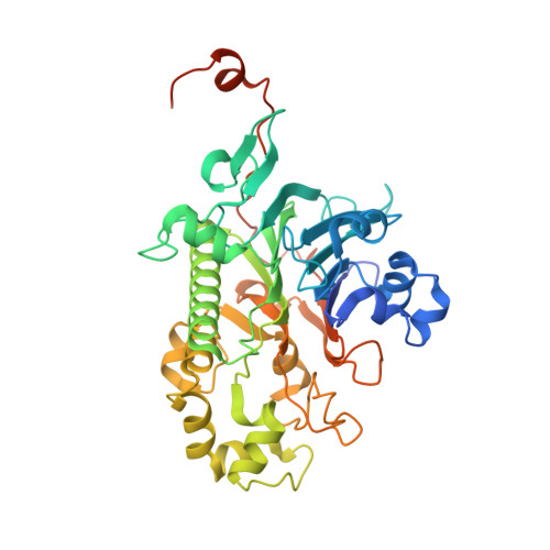
    Topoisomerase 1 (Top1) incises DNA containing ribonucleotides to generate complex DNA lesions that are resolved by APE2 (Apn2 in yeast). How Apn2 engages and processes this DNA damage is unclear. Here, we report X-ray crystal structures and biochemical analysis of Apn2-DNA complexes to demonstrate how Apn2 frays and cleaves 3' DNA termini via a wedging mechanism that facilitates 1-6 nucleotide endonucleolytic cleavages. APN2 deletion and DNA-wedge mutant Saccharomyces cerevisiae strains display mutator phenotypes, cell growth defects, and sensitivity to genotoxic stress in a ribonucleotide excision repair (RER)-defective background harboring a high density of Top1-incised ribonucleotides. Our data implicate a wedge-and-cut mechanism underpinning the broad-specificity Apn2 nuclease activity that mitigates mutagenic and genome instability phenotypes caused by Top1 incision at genomic ribonucleotides incorporated by DNA polymerase epsilon.


  • Organizational Affiliation
    • Genome Integrity and Structural Biology Laboratory, National Institute of Environmental Health Sciences, National Institutes of Health, US Department of Health and Human Services, Research Triangle Park, NC 27709, USA.

Macromolecule Content 

  • Total Structure Weight: 47.45 kDa 
  • Atom Count: 3,246 
  • Modeled Residue Count: 379 
  • Deposited Residue Count: 413 
  • Unique protein chains: 1

Macromolecules

Find similar proteins by:|  3D Structure
Entity ID: 1
MoleculeChains  Sequence LengthOrganismDetailsImage
DNA-(apurinic or apyrimidinic site) endonuclease 2413Saccharomyces cerevisiaeMutation(s): 0 
Gene Names: APN2ETH1YBL019WYBL0443
EC: 3.1 (PDB Primary Data), 3.1.11.2 (UniProt)
UniProt
Find proteins for P38207 (Saccharomyces cerevisiae (strain ATCC 204508 / S288c))
Explore P38207 
Go to UniProtKB:  P38207
Entity Groups
Sequence Clusters30% Identity50% Identity70% Identity90% Identity95% Identity100% Identity
UniProt GroupP38207
Sequence Annotations
Expand
Reference Sequence

Experimental Data & Validation

Experimental Data

  • Method: X-RAY DIFFRACTION
  • Resolution: 1.99 Å
  • R-Value Free:  0.198 (Depositor), 0.198 (DCC) 
  • R-Value Work:  0.163 (Depositor), 0.164 (DCC) 
  • R-Value Observed: 0.164 (Depositor) 
Space Group: C 1 2 1
Unit Cell:
Length ( Å )Angle ( ˚ )
a = 171.412α = 90
b = 46.948β = 104.213
c = 51.532γ = 90
Software Package:
Software NamePurpose
PHENIXrefinement
HKL-2000data reduction
HKL-2000data scaling
PHENIXphasing
Cootmodel building

Structure Validation

View Full Validation Report


Entry History 

& Funding Information

Deposition Data


Funding OrganizationLocationGrant Number
National Institutes of Health/National Institute of Environmental Health Sciences (NIH/NIEHS)United States1Z01ES102765

Revision History  (Full details and data files)

  • Version 1.0: 2022-09-28
    Type: Initial release
  • Version 1.1: 2022-10-19
    Changes: Database references
  • Version 1.2: 2023-10-18
    Changes: Data collection, Refinement description