7LZD | pdb_00007lzd

Crystal Structure of SETD2 bound to Compound 35


Experimental Data Snapshot

  • Method: X-RAY DIFFRACTION
  • Resolution: 1.80 Å
  • R-Value Free: 
    0.218 (Depositor), 0.227 (DCC) 
  • R-Value Work: 
    0.193 (Depositor), 0.204 (DCC) 
  • R-Value Observed: 
    0.194 (Depositor) 

Starting Model: experimental
View more details

wwPDB Validation 3D Report Full Report

Validation slider image for 7LZD

Ligand Structure Quality Assessment 


This is version 1.2 of the entry. See complete history

Literature

Discovery of a First-in-Class Inhibitor of the Histone Methyltransferase SETD2 Suitable for Preclinical Studies.

Lampe, J.W.Alford, J.S.Boriak-Sjodin, P.A.Brach, D.Cosmopoulos, K.Duncan, K.W.Eckley, S.T.Foley, M.A.Harvey, D.M.Motwani, V.Munchhof, M.J.Raimondi, A.Riera, T.V.Tang, C.Thomenius, M.J.Totman, J.Farrow, N.A.

(2021) ACS Med Chem Lett 12: 1539-1545

  • DOI: https://doi.org/10.1021/acsmedchemlett.1c00272
  • Primary Citation Related Structures: 
    7LZB, 7LZD, 7LZF

  • PubMed Abstract: 

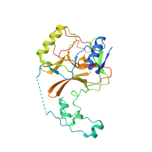
    SET domain-containing protein 2 (SETD2), a histone methyltransferase, has been identified as a target of interest in certain hematological malignancies, including multiple myeloma. This account details the discovery of EPZ-719 , a novel and potent SETD2 inhibitor with a high selectivity over other histone methyltransferases. A screening campaign of the Epizyme proprietary histone methyltransferase-biased library identified potential leads based on a 2-amidoindole core. Structure-based drug design (SBDD) and drug metabolism/pharmacokinetics (DMPK) optimization resulted in EPZ-719 , an attractive tool compound for the interrogation of SETD2 biology that enables in vivo target validation studies.


  • Organizational Affiliation
    • Epizyme Inc., 400 Technology Square, Fourth Floor, Cambridge, Massachusetts 02139, United States.

Macromolecule Content 

  • Total Structure Weight: 33.58 kDa 
  • Atom Count: 2,177 
  • Modeled Residue Count: 237 
  • Deposited Residue Count: 278 
  • Unique protein chains: 1

Macromolecules

Find similar proteins by:|  3D Structure
Entity ID: 1
MoleculeChains  Sequence LengthOrganismDetailsImage
Histone-lysine N-methyltransferase SETD2278Homo sapiensMutation(s): 0 
Gene Names: SETD2HIF1HYPBKIAA1732KMT3ASET2HSPC069
EC: 2.1.1.359 (PDB Primary Data), 2.1.1 (PDB Primary Data)
UniProt & NIH Common Fund Data Resources
Find proteins for Q9BYW2 (Homo sapiens)
Explore Q9BYW2 
Go to UniProtKB:  Q9BYW2
PHAROS:  Q9BYW2
GTEx:  ENSG00000181555 
Entity Groups
Sequence Clusters30% Identity50% Identity70% Identity90% Identity95% Identity100% Identity
UniProt GroupQ9BYW2
Sequence Annotations
Expand
Reference Sequence

Small Molecules

Ligands 5 Unique
IDChains Name / Formula / InChI Key2D Diagram3D Interactions
SAM

Query on SAM


Download:Ideal Coordinates CCD File
E [auth A]S-ADENOSYLMETHIONINE
C15 H22 N6 O5 S
MEFKEPWMEQBLKI-FCKMPRQPSA-N
YHY
(Subject of Investigation/LOI)

Query on YHY


Download:Ideal Coordinates CCD File
F [auth A]N-{3-[4-(dimethylamino)piperidin-1-yl]phenyl}-4-fluoro-7-methyl-1H-indole-2-carboxamide
C23 H27 F N4 O
GBIAQCRLFDZGOQ-UHFFFAOYSA-N
DMS

Query on DMS


Download:Ideal Coordinates CCD File
P [auth A]DIMETHYL SULFOXIDE
C2 H6 O S
IAZDPXIOMUYVGZ-UHFFFAOYSA-N
ZN

Query on ZN


Download:Ideal Coordinates CCD File
B [auth A],
C [auth A],
D [auth A]
ZINC ION
Zn
PTFCDOFLOPIGGS-UHFFFAOYSA-N
EDO

Query on EDO


Download:Ideal Coordinates CCD File
G [auth A]
H [auth A]
I [auth A]
J [auth A]
K [auth A]
G [auth A],
H [auth A],
I [auth A],
J [auth A],
K [auth A],
L [auth A],
M [auth A],
N [auth A],
O [auth A]
1,2-ETHANEDIOL
C2 H6 O2
LYCAIKOWRPUZTN-UHFFFAOYSA-N
Binding Affinity Annotations 
IDSourceBinding Affinity
YHY BindingDB:  7LZD IC50: min: 18, max: 235 (nM) from 2 assay(s)

Experimental Data & Validation

Experimental Data

  • Method: X-RAY DIFFRACTION
  • Resolution: 1.80 Å
  • R-Value Free:  0.218 (Depositor), 0.227 (DCC) 
  • R-Value Work:  0.193 (Depositor), 0.204 (DCC) 
  • R-Value Observed: 0.194 (Depositor) 
Space Group: P 21 21 21
Unit Cell:
Length ( Å )Angle ( ˚ )
a = 52.231α = 90
b = 76.381β = 90
c = 76.434γ = 90
Software Package:
Software NamePurpose
REFMACrefinement
PDB_EXTRACTdata extraction
HKL-3000data reduction
HKL-3000data scaling

Structure Validation

View Full Validation Report


Ligand Structure Quality Assessment 


Entry History 

Deposition Data

Revision History  (Full details and data files)

  • Version 1.0: 2021-09-22
    Type: Initial release
  • Version 1.1: 2021-11-03
    Changes: Database references
  • Version 1.2: 2023-10-18
    Changes: Data collection, Refinement description