7LSA | pdb_00007lsa

Ruminococcus bromii Amy12 with maltoheptaose


Experimental Data Snapshot

  • Method: X-RAY DIFFRACTION
  • Resolution: 1.76 Å
  • R-Value Free: 
    0.206 (Depositor), 0.218 (DCC) 
  • R-Value Work: 
    0.159 (Depositor), 0.171 (DCC) 
  • R-Value Observed: 
    0.160 (Depositor) 

Starting Model: experimental
View more details

wwPDB Validation 3D Report Full Report

Validation slider image for 7LSA

This is version 1.1 of the entry. See complete history

Literature

Structure and substrate recognition by the Ruminococcus bromii amylosome pullulanases.

Cockburn, D.W.Kibler, R.Brown, H.A.Duvall, R.Morais, S.Bayer, E.Koropatkin, N.M.

(2021) J Struct Biol 213: 107765-107765

  • DOI: https://doi.org/10.1016/j.jsb.2021.107765
  • Primary Citation Related Structures: 
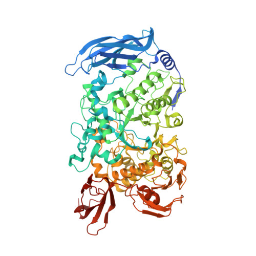
    7LSA, 7LSR, 7LST, 7LSU

  • PubMed Abstract: 

    Pullulanases are glycoside hydrolase family 13 (GH13) enzymes that target α1,6 glucosidic linkages within starch and aid in the degradation of the α1,4- and α1,6- linked glucans pullulan, glycogen and amylopectin. The human gut bacterium Ruminococcus bromii synthesizes two extracellular pullulanases, Amy10 and Amy12, that are incorporated into the multiprotein amylosome complex that enables the digestion of granular resistant starch from the diet. Here we provide a comparative biochemical analysis of these pullulanases and the x-ray crystal structures of the wild type and the nucleophile mutant D392A of Amy12 complexed with maltoheptaose and 6 3 -α-D glucosyl-maltotriose. While Amy10 displays higher catalytic efficiency on pullulan and cleaves only α1,6 linkages, Amy12 has some activity on α1,4 linkages suggesting that these enzymes are not redundant within the amylosome. Our structures of Amy12 include a mucin-binding protein (MucBP) domain that follows the C-domain of the GH13 fold, an atypical feature of these enzymes. The wild type Amy12 structure with maltoheptaose captured two oligosaccharides in the active site arranged as expected following catalysis of an α1,6 branch point in amylopectin. The nucleophile mutant D392A complexed with maltoheptaose or 6 3 -α-D glucosyl-maltotriose captured β-glucose at the reducing end in the -1 subsite, facilitated by the truncation of the active site aspartate and stabilized by stacking with Y279. The core interface between the co-crystallized ligands and Amy12 occurs within the -2 through + 1 subsites, which may allow for flexible recognition of α1,6 linkages within a variety of starch structures.


  • Organizational Affiliation
    • Department of Microbiology and Immunology, University of Michigan Medical School, Ann Arbor, MI 48109, United States.

Macromolecule Content 

  • Total Structure Weight: 89.39 kDa 
  • Atom Count: 6,748 
  • Modeled Residue Count: 760 
  • Deposited Residue Count: 797 
  • Unique protein chains: 1

Macromolecules

Find similar proteins by:|  3D Structure
Entity ID: 1
MoleculeChains  Sequence LengthOrganismDetailsImage
Pullulanase797Ruminococcus bromiiMutation(s): 0 
Gene Names: pulA_2RBL236_00821
EC: 3.2.1.41

Oligosaccharides

Help  
Entity ID: 2
MoleculeChains Length2D Diagram GlycosylationD Interactions
alpha-D-glucopyranose-(1-4)-alpha-D-glucopyranose-(1-4)-alpha-D-glucopyranose-(1-4)-alpha-D-glucopyranose-(1-4)-alpha-D-glucopyranoseB [auth C]5N/A
Glycosylation Resources
GlyTouCan: G50146AM
GlyCosmos: G50146AM
Entity ID: 3
MoleculeChains Length2D Diagram GlycosylationD Interactions
alpha-D-glucopyranose-(1-4)-alpha-D-glucopyranose-(1-4)-alpha-D-glucopyranoseC [auth D]3N/A
Glycosylation Resources
GlyTouCan: G96370VA
GlyCosmos: G96370VA
GlyGen: G96370VA

Small Molecules

Ligands 3 Unique
IDChains Name / Formula / InChI Key2D Diagram3D Interactions
PEG

Query on PEG


Download:Ideal Coordinates CCD File
L [auth A]DI(HYDROXYETHYL)ETHER
C4 H10 O3
MTHSVFCYNBDYFN-UHFFFAOYSA-N
GOL

Query on GOL


Download:Ideal Coordinates CCD File
D [auth A]
E [auth A]
F [auth A]
G [auth A]
H [auth A]
D [auth A],
E [auth A],
F [auth A],
G [auth A],
H [auth A],
I [auth A],
J [auth A],
K [auth A]
GLYCEROL
C3 H8 O3
PEDCQBHIVMGVHV-UHFFFAOYSA-N
CA

Query on CA


Download:Ideal Coordinates CCD File
M [auth A]CALCIUM ION
Ca
BHPQYMZQTOCNFJ-UHFFFAOYSA-N

Biologically Interesting Molecules (External Reference) 

2 Unique

Experimental Data & Validation

Experimental Data

  • Method: X-RAY DIFFRACTION
  • Resolution: 1.76 Å
  • R-Value Free:  0.206 (Depositor), 0.218 (DCC) 
  • R-Value Work:  0.159 (Depositor), 0.171 (DCC) 
  • R-Value Observed: 0.160 (Depositor) 
Space Group: P 21 21 21
Unit Cell:
Length ( Å )Angle ( ˚ )
a = 46.977α = 90
b = 97.96β = 90
c = 167.089γ = 90
Software Package:
Software NamePurpose
xia2data scaling
PHASERphasing
REFMACrefinement
PDB_EXTRACTdata extraction
xia2data reduction

Structure Validation

View Full Validation Report


Entry History 

& Funding Information

Deposition Data


Funding OrganizationLocationGrant Number
United States - Israel Binational Science Foundation (BSF)United States--

Revision History  (Full details and data files)

  • Version 1.0: 2021-07-14
    Type: Initial release
  • Version 1.1: 2023-10-18
    Changes: Data collection, Database references, Refinement description