7LNH | pdb_00007lnh

S-adenosylmethionine synthetase co-crystallized with UppNHp


Experimental Data Snapshot

  • Method: X-RAY DIFFRACTION
  • Resolution: 2.50 Å
  • R-Value Free: 
    0.246 (Depositor), 0.249 (DCC) 
  • R-Value Work: 
    0.219 (Depositor), 0.222 (DCC) 
  • R-Value Observed: 
    0.221 (Depositor) 

Starting Model: experimental
View more details

wwPDB Validation 3D Report Full Report

Validation slider image for 7LNH

Ligand Structure Quality Assessment 


This is version 1.1 of the entry. See complete history

Literature

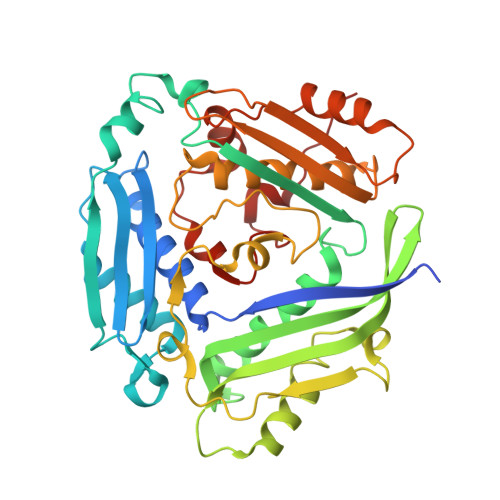
Substrate Dynamics Contribute to Enzymatic Specificity in Human and Bacterial Methionine Adenosyltransferases.

Gade, M.Tan, L.L.Damry, A.M.Sandhu, M.Brock, J.S.Delaney, A.Villar-Briones, A.Jackson, C.J.Laurino, P.

(2021) JACS Au 1: 2349-2360

  • DOI: https://doi.org/10.1021/jacsau.1c00464
  • Primary Citation Related Structures: 
    7LL3, 7LNH, 7LNN, 7LO2, 7LOO, 7LOW, 7LOZ

  • PubMed Abstract: 

    Protein conformational changes can facilitate the binding of noncognate substrates and underlying promiscuous activities. However, the contribution of substrate conformational dynamics to this process is comparatively poorly understood. Here, we analyze human (hMAT2A) and Escherichia coli (eMAT) methionine adenosyltransferases that have identical active sites but different substrate specificity. In the promiscuous hMAT2A, noncognate substrates bind in a stable conformation to allow catalysis. In contrast, noncognate substrates sample stable productive binding modes less frequently in eMAT owing to altered mobility in the enzyme active site. Different cellular concentrations of substrates likely drove the evolutionary divergence of substrate specificity in these orthologues. The observation of catalytic promiscuity in hMAT2A led to the detection of a new human metabolite, methyl thioguanosine, that is produced at elevated levels in a cancer cell line. This work establishes that identical active sites can result in different substrate specificity owing to the effects of substrate and enzyme dynamics.


  • Organizational Affiliation
    • Protein Engineering and Evolution Unit, Okinawa Institute of Science and Technology Graduate University, 1919-1 Tancha, Onna 904-0495, Okinawa, Japan.

Macromolecule Content 

  • Total Structure Weight: 88.25 kDa 
  • Atom Count: 6,146 
  • Modeled Residue Count: 761 
  • Deposited Residue Count: 790 
  • Unique protein chains: 1

Macromolecules

Find similar proteins by:|  3D Structure
Entity ID: 1
MoleculeChains  Sequence LengthOrganismDetailsImage
S-adenosylmethionine synthase isoform type-2
A, B
395Homo sapiensMutation(s): 0 
Gene Names: MAT2AAMS2MATA2
EC: 2.5.1.6
UniProt & NIH Common Fund Data Resources
Find proteins for P31153 (Homo sapiens)
Explore P31153 
Go to UniProtKB:  P31153
PHAROS:  P31153
GTEx:  ENSG00000168906 
Entity Groups
Sequence Clusters30% Identity50% Identity70% Identity90% Identity95% Identity100% Identity
UniProt GroupP31153
Sequence Annotations
Expand
Reference Sequence

Small Molecules

Ligands 2 Unique
IDChains Name / Formula / InChI Key2D Diagram3D Interactions
YQP
(Subject of Investigation/LOI)

Query on YQP


Download:Ideal Coordinates CCD File
C [auth A],
E [auth B]
(2~{S})-2-azanyl-4-[[(2~{S},3~{S},4~{R},5~{R})-5-[2,4-bis(oxidanylidene)pyrimidin-1-yl]-3,4-bis(oxidanyl)oxolan-2-yl]methyl-methyl-$l^{3}-sulfanyl]butanoic acid
C14 H21 N3 O7 S
HBPDMWXHIXRRKQ-GGUSWIIGSA-N
EDO

Query on EDO


Download:Ideal Coordinates CCD File
D [auth B]1,2-ETHANEDIOL
C2 H6 O2
LYCAIKOWRPUZTN-UHFFFAOYSA-N

Experimental Data & Validation

Experimental Data

  • Method: X-RAY DIFFRACTION
  • Resolution: 2.50 Å
  • R-Value Free:  0.246 (Depositor), 0.249 (DCC) 
  • R-Value Work:  0.219 (Depositor), 0.222 (DCC) 
  • R-Value Observed: 0.221 (Depositor) 
Space Group: P 63 2 2
Unit Cell:
Length ( Å )Angle ( ˚ )
a = 144.191α = 90
b = 144.191β = 90
c = 188.196γ = 120
Software Package:
Software NamePurpose
PHENIXrefinement
XDSdata reduction
Aimlessdata scaling
PHASERphasing

Structure Validation

View Full Validation Report


Ligand Structure Quality Assessment 


Entry History 

Deposition Data

Revision History  (Full details and data files)

  • Version 1.0: 2022-03-02
    Type: Initial release
  • Version 1.1: 2023-10-18
    Changes: Data collection, Refinement description