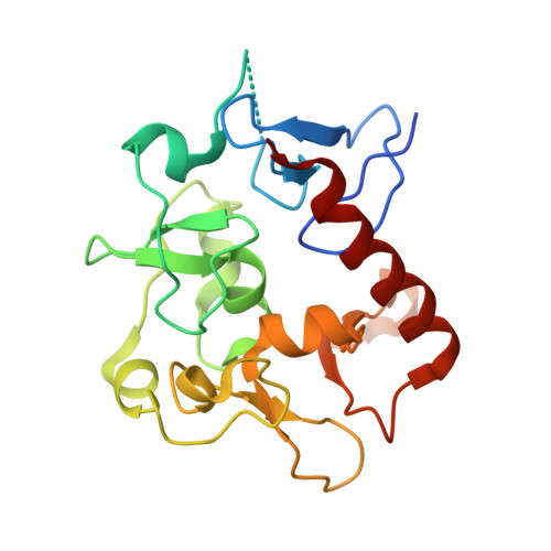
7D86

Crystal Structure of zebrafishPHF14-PZP


Experimental Data Snapshot

  • Method: X-RAY DIFFRACTION
  • Resolution: 1.84 Å
  • R-Value Free: 0.225 
  • R-Value Work: 0.174 
  • R-Value Observed: 0.179 

wwPDB Validation   3D Report Full Report


This is version 1.2 of the entry. See complete history


Literature

Molecular basis for bipartite recognition of histone H3 by the PZP domain of PHF14.

Zheng, S.Bi, Y.Chen, H.Gong, B.Jia, S.Li, H.

(2021) Nucleic Acids Res 49: 8961-8973

  • DOI: https://doi.org/10.1093/nar/gkab670
  • Primary Citation of Related Structures:  
    7D86, 7D87, 7D8A

  • PubMed Abstract: 

    Histone recognition constitutes a key epigenetic mechanism in gene regulation and cell fate decision. PHF14 is a conserved multi-PHD finger protein that has been implicated in organ development, tissue homeostasis, and tumorigenesis. Here we show that PHF14 reads unmodified histone H3(1-34) through an integrated PHD1-ZnK-PHD2 cassette (PHF14PZP). Our binding, structural and HDX-MS analyses revealed a feature of bipartite recognition, in which PHF14PZP utilizes two distinct surfaces for concurrent yet separable engagement of segments H3-Nter (e.g. 1-15) and H3-middle (e.g. 14-34) of H3(1-34). Structural studies revealed a novel histone H3 binding mode by PHD1 of PHF14PZP, in which a PHF14-unique insertion loop but not the core β-strands of a PHD finger dominates H3K4 readout. Binding studies showed that H3-PHF14PZP engagement is sensitive to modifications occurring to H3 R2, T3, K4, R8 and K23 but not K9 and K27, suggesting multiple layers of modification switch. Collectively, our work calls attention to PHF14 as a 'ground' state (unmodified) H3(1-34) reader that can be negatively regulated by active marks, thus providing molecular insights into a repressive function of PHF14 and its derepression.


  • Organizational Affiliation

    MOE Key Laboratory of Protein Sciences, Beijing Frontier Research Center for Biological Structure, Beijing Advanced Innovation Center for Structural Biology, Department of Basic Medical Sciences, School of Medicine, Tsinghua University, Beijing 100084, China.


Macromolecules
Find similar proteins by:  (by identity cutoff)  |  3D Structure
Entity ID: 1
MoleculeChains Sequence LengthOrganismDetailsImage
PHD finger protein 14211Danio rerioMutation(s): 0 
Gene Names: phf14
UniProt
Find proteins for A0A286Y9D1 (Danio rerio)
Explore A0A286Y9D1 
Go to UniProtKB:  A0A286Y9D1
Entity Groups  
Sequence Clusters30% Identity50% Identity70% Identity90% Identity95% Identity100% Identity
UniProt GroupA0A286Y9D1
Sequence Annotations
Expand
  • Reference Sequence
Small Molecules
Ligands 3 Unique
IDChains Name / Formula / InChI Key2D Diagram3D Interactions
ZN
Query on ZN

Download Ideal Coordinates CCD File 
B [auth A],
C [auth A],
D [auth A],
E [auth A],
F [auth A]
ZINC ION
Zn
PTFCDOFLOPIGGS-UHFFFAOYSA-N
EDO
Query on EDO

Download Ideal Coordinates CCD File 
H [auth A]1,2-ETHANEDIOL
C2 H6 O2
LYCAIKOWRPUZTN-UHFFFAOYSA-N
MG
Query on MG

Download Ideal Coordinates CCD File 
G [auth A]MAGNESIUM ION
Mg
JLVVSXFLKOJNIY-UHFFFAOYSA-N
Experimental Data & Validation

Experimental Data

  • Method: X-RAY DIFFRACTION
  • Resolution: 1.84 Å
  • R-Value Free: 0.225 
  • R-Value Work: 0.174 
  • R-Value Observed: 0.179 
  • Space Group: P 1 21 1
Unit Cell:
Length ( Å )Angle ( ˚ )
a = 42.193α = 90
b = 41.907β = 98.95
c = 52.878γ = 90
Software Package:
Software NamePurpose
HKL-2000data scaling
PHENIXrefinement
PDB_EXTRACTdata extraction
HKL-2000data reduction
MOLREPphasing

Structure Validation

View Full Validation Report


Entry History & Funding Information

Deposition Data


Funding OrganizationLocationGrant Number
National Natural Science Foundation of China (NSFC)China--

Revision History  (Full details and data files)

  • Version 1.0: 2021-07-28
    Type: Initial release
  • Version 1.1: 2022-02-09
    Changes: Database references, Structure summary
  • Version 1.2: 2024-05-29
    Changes: Data collection