7CJT | pdb_00007cjt

Crystal Structure of SETDB1 Tudor domain in complexed with (R,R)-59


Experimental Data Snapshot

  • Method: X-RAY DIFFRACTION
  • Resolution: 2.47 Å
  • R-Value Free: 
    0.276 (Depositor), 0.279 (DCC) 
  • R-Value Work: 
    0.204 (Depositor), 0.207 (DCC) 
  • R-Value Observed: 
    0.208 (Depositor) 

Starting Model: experimental
View more details

wwPDB Validation 3D Report Full Report

Validation slider image for 7CJT

Ligand Structure Quality Assessment 


This is version 1.1 of the entry. See complete history

Literature

Structure-Guided Discovery of a Potent and Selective Cell-Active Inhibitor of SETDB1 Tudor Domain.

Guo, Y.Mao, X.Xiong, L.Xia, A.You, J.Lin, G.Wu, C.Huang, L.Wang, Y.Yang, S.

(2021) Angew Chem Int Ed Engl 60: 8760-8765

  • DOI: https://doi.org/10.1002/anie.202017200
  • Primary Citation Related Structures: 
    7C9N, 7CAJ, 7CD9, 7CJT

  • PubMed Abstract: 

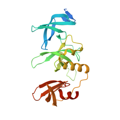
    SET domain bifurcated protein 1 (SETDB1) is a histone lysine methyltransferase that promotes the silencing of some tumour suppressor genes and is overexpressed in many cancers. SETDB1 contains a unique tandem tudor domain (TTD) that recognizes histone H3 sequences containing both methylated and acetylated lysines. Beginning with the identification of a hit compound (Cpd1), we discovered the first potent and selective small molecule SETDB1-TTD inhibitor (R,R)-59 through stepwise structure-guided optimization. (R,R)-59 showed a K D value of 0.088±0.045 μM in the ITC assay. The high potency of (R,R)-59 was well explained by the cocrystal structure of the (R,R)-59-TTD complex. (R,R)-59 is an endogenous binder competitive inhibitor. Evidence has also demonstrated its cellular target engagement. Interestingly, the enantiomer (S,S)-59 did not show activity in all the assays, highlighting the potential of (R,R)-59 as a tool compound in exploring the biological functions of SETDB1-TTD.


  • Organizational Affiliation
    • State Key Laboratory of Biotherapy and Cancer Center, West China Hospital, Sichuan University, Chengdu, Sichuan, 610041, P. R. China.

Macromolecule Content 

  • Total Structure Weight: 112.33 kDa 
  • Atom Count: 6,993 
  • Modeled Residue Count: 854 
  • Deposited Residue Count: 960 
  • Unique protein chains: 1

Macromolecules

Find similar proteins by:|  3D Structure
Entity ID: 1
MoleculeChains  Sequence LengthOrganismDetailsImage
Histone-lysine N-methyltransferase SETDB1A [auth D],
B [auth A],
C [auth B],
D [auth C]
240Homo sapiensMutation(s): 0 
Gene Names: SETDB1ESETKIAA0067KMT1E
EC: 2.1.1 (PDB Primary Data), 2.1.1.366 (UniProt)
UniProt & NIH Common Fund Data Resources
Find proteins for Q15047 (Homo sapiens)
Explore Q15047 
Go to UniProtKB:  Q15047
PHAROS:  Q15047
GTEx:  ENSG00000143379 
Entity Groups
Sequence Clusters30% Identity50% Identity70% Identity90% Identity95% Identity100% Identity
UniProt GroupQ15047
Sequence Annotations
Expand
Reference Sequence

Small Molecules

Binding Affinity Annotations 
IDSourceBinding Affinity
G09 BindingDB:  7CJT Kd: 80 (nM) from 1 assay(s)
IC50: min: 7.50e+5, max: 9.30e+5 (nM) from 2 assay(s)

Experimental Data & Validation

Experimental Data

  • Method: X-RAY DIFFRACTION
  • Resolution: 2.47 Å
  • R-Value Free:  0.276 (Depositor), 0.279 (DCC) 
  • R-Value Work:  0.204 (Depositor), 0.207 (DCC) 
  • R-Value Observed: 0.208 (Depositor) 
Space Group: P 1 21 1
Unit Cell:
Length ( Å )Angle ( ˚ )
a = 83.335α = 90
b = 69.214β = 98.93
c = 110.124γ = 90
Software Package:
Software NamePurpose
SCALAdata scaling
PHENIXrefinement
PDB_EXTRACTdata extraction
XDSdata reduction
PHASERphasing

Structure Validation

View Full Validation Report


Ligand Structure Quality Assessment 


Entry History 

& Funding Information

Deposition Data


Funding OrganizationLocationGrant Number
National Natural Science Foundation of China (NSFC)China81930125

Revision History  (Full details and data files)

  • Version 1.0: 2021-04-14
    Type: Initial release
  • Version 1.1: 2023-11-29
    Changes: Data collection, Database references, Refinement description