7B6W

Crystal structure of the human alpha1B adrenergic receptor in complex with inverse agonist (+)-cyclazosin


Experimental Data Snapshot

  • Method: X-RAY DIFFRACTION
  • Resolution: 2.87 Å
  • R-Value Free: 0.317 
  • R-Value Work: 0.289 

wwPDB Validation   3D Report Full Report


Ligand Structure Quality Assessment 


This is version 2.0 of the entry. See complete history


Literature

Crystal structure of the alpha 1B -adrenergic receptor reveals molecular determinants of selective ligand recognition.

Deluigi, M.Morstein, L.Schuster, M.Klenk, C.Merklinger, L.Cridge, R.R.de Zhang, L.A.Klipp, A.Vacca, S.Vaid, T.M.Mittl, P.R.E.Egloff, P.Eberle, S.A.Zerbe, O.Chalmers, D.K.Scott, D.J.Pluckthun, A.

(2022) Nat Commun 13: 382-382

  • DOI: https://doi.org/10.1038/s41467-021-27911-3
  • Primary Citation of Related Structures:  
    7B6W

  • PubMed Abstract: 

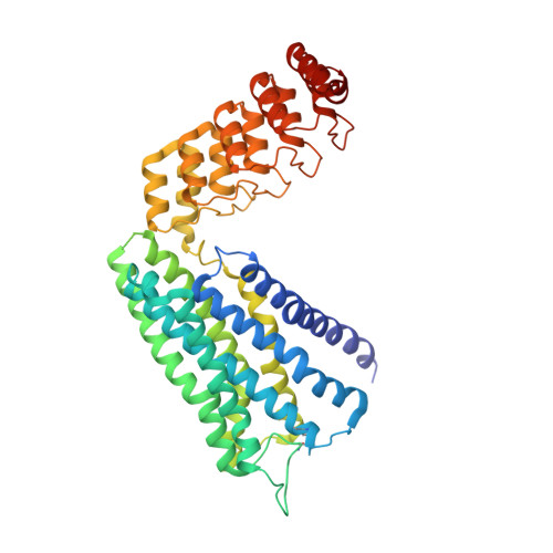
    α-adrenergic receptors (αARs) are G protein-coupled receptors that regulate vital functions of the cardiovascular and nervous systems. The therapeutic potential of αARs, however, is largely unexploited and hampered by the scarcity of subtype-selective ligands. Moreover, several aminergic drugs either show off-target binding to αARs or fail to interact with the desired subtype. Here, we report the crystal structure of human α 1B AR bound to the inverse agonist (+)-cyclazosin, enabled by the fusion to a DARPin crystallization chaperone. The α 1B AR structure allows the identification of two unique secondary binding pockets. By structural comparison of α 1B AR with α 2 ARs, and by constructing α 1B AR-α 2C AR chimeras, we identify residues 3.29 and 6.55 as key determinants of ligand selectivity. Our findings provide a basis for discovery of α 1B AR-selective ligands and may guide the optimization of aminergic drugs to prevent off-target binding to αARs, or to elicit a selective interaction with the desired subtype.


  • Organizational Affiliation

    Department of Biochemistry, University of Zurich, Winterthurerstrasse 190, CH-8057, Zurich, Switzerland.


Macromolecules
Find similar proteins by:  (by identity cutoff)  |  3D Structure
Entity ID: 1
MoleculeChains Sequence LengthOrganismDetailsImage
Alpha-1B adrenergic receptor,alpha1B adrenergic receptor,Alpha-1B adrenergic receptor,alpha1B adrenergic receptor,Alpha-1B adrenergic receptor,alpha1B adrenergic receptor,Alpha-1B adrenergic receptor,alpha1B adrenergic receptorA [auth AAA]459Homo sapiensMutation(s): 0 
Gene Names: ADRA1B
Entity Groups  
Sequence Clusters30% Identity50% Identity70% Identity90% Identity95% Identity100% Identity
Sequence Annotations
Expand
  • Reference Sequence
Small Molecules
Ligands 1 Unique
IDChains Name / Formula / InChI Key2D Diagram3D Interactions
T0B (Subject of Investigation/LOI)
Query on T0B

Download Ideal Coordinates CCD File 
B [auth AAA][(4~{a}~{R},8~{a}~{S})-4-(4-azanyl-6,7-dimethoxy-quinazolin-2-yl)-2,3,4~{a},5,6,7,8,8~{a}-octahydroquinoxalin-1-yl]-(furan-2-yl)methanone
C23 H27 N5 O4
XBRXTUGRUXGBPX-DLBZAZTESA-N
Experimental Data & Validation

Experimental Data

  • Method: X-RAY DIFFRACTION
  • Resolution: 2.87 Å
  • R-Value Free: 0.317 
  • R-Value Work: 0.289 
  • Space Group: P 21 21 21
Unit Cell:
Length ( Å )Angle ( ˚ )
a = 67.883α = 90
b = 76.415β = 90
c = 151.634γ = 90
Software Package:
Software NamePurpose
REFMACrefinement
XDSdata reduction
Aimlessdata scaling
PHASERphasing

Structure Validation

View Full Validation Report


Ligand Structure Quality Assessment 


Entry History & Funding Information

Deposition Data


Funding OrganizationLocationGrant Number
Swiss National Science FoundationSwitzerland31003A_182334

Revision History  (Full details and data files)

  • Version 1.0: 2022-01-12
    Type: Initial release
  • Version 1.1: 2022-02-02
    Changes: Database references
  • Version 1.2: 2024-01-31
    Changes: Data collection, Derived calculations, Refinement description
  • Version 2.0: 2024-05-01
    Changes: Atomic model, Data collection, Derived calculations, Structure summary