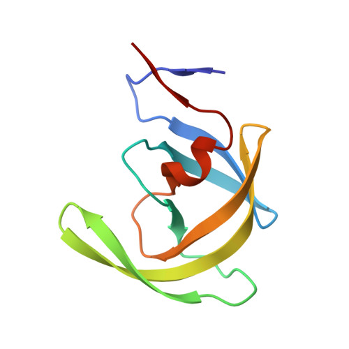
6PJL

HIV-1 Protease NL4-3 WT in Complex with LR3-95


Experimental Data Snapshot

  • Method: X-RAY DIFFRACTION
  • Resolution: 1.99 Å
  • R-Value Free: 0.231 
  • R-Value Work: 0.200 
  • R-Value Observed: 0.202 

Starting Model: experimental
View more details

wwPDB Validation   3D Report Full Report


Ligand Structure Quality Assessment 


This is version 1.3 of the entry. See complete history


Literature

Structural Analysis of Potent Hybrid HIV-1 Protease Inhibitors Containing Bis-tetrahydrofuran in a Pseudosymmetric Dipeptide Isostere.

Rusere, L.N.Lockbaum, G.J.Henes, M.Lee, S.K.Spielvogel, E.Rao, D.N.Kosovrasti, K.Nalivaika, E.A.Swanstrom, R.Kurt Yilmaz, N.Schiffer, C.A.Ali, A.

(2020) J Med Chem 63: 8296-8313

  • DOI: https://doi.org/10.1021/acs.jmedchem.0c00529
  • Primary Citation of Related Structures:  
    6PJB, 6PJC, 6PJD, 6PJE, 6PJF, 6PJG, 6PJH, 6PJI, 6PJK, 6PJL, 6PJM, 6PJN, 6PJO

  • PubMed Abstract: 

    The design, synthesis, and X-ray structural analysis of hybrid HIV-1 protease inhibitors (PIs) containing bis-tetrahydrofuran (bis-THF) in a pseudo-C 2 -symmetric dipeptide isostere are described. A series of PIs were synthesized by incorporating bis-THF of darunavir on either side of the Phe-Phe isostere of lopinavir in combination with hydrophobic amino acids on the opposite P2/P2' position. Structure-activity relationship studies indicated that the bis-THF moiety can be attached at either the P2 or P2' position without significantly affecting potency. However, the group on the opposite P2/P2' position had a dramatic effect on potency depending on the size and shape of the side chain. Cocrystal structures of inhibitors with wild-type HIV-1 protease revealed that the bis-THF moiety retained similar interactions as observed in the darunavir-protease complex regardless of the position on the Phe-Phe isostere. Analyses of cocrystal structures and molecular dynamics simulations provide insights into optimizing HIV-1 PIs containing bis-THF in non-sulfonamide dipeptide isosteres.


  • Organizational Affiliation

    Department of Biochemistry and Molecular Pharmacology, University of Massachusetts Medical School, Worcester, Massachusetts 01605, United States.


Macromolecules
Find similar proteins by:  (by identity cutoff)  |  3D Structure
Entity ID: 1
MoleculeChains Sequence LengthOrganismDetailsImage
Protease NL4-3A [auth B],
B [auth A]
99Human immunodeficiency virus 1Mutation(s): 1 
Gene Names: pol
UniProt
Find proteins for P12497 (Human immunodeficiency virus type 1 group M subtype B (isolate NY5))
Explore P12497 
Go to UniProtKB:  P12497
Entity Groups  
Sequence Clusters30% Identity50% Identity70% Identity90% Identity95% Identity100% Identity
UniProt GroupP12497
Sequence Annotations
Expand
  • Reference Sequence
Experimental Data & Validation

Experimental Data

  • Method: X-RAY DIFFRACTION
  • Resolution: 1.99 Å
  • R-Value Free: 0.231 
  • R-Value Work: 0.200 
  • R-Value Observed: 0.202 
  • Space Group: P 21 21 21
Unit Cell:
Length ( Å )Angle ( ˚ )
a = 51.455α = 90
b = 58.187β = 90
c = 61.71γ = 90
Software Package:
Software NamePurpose
HKL-2000data scaling
PHASERphasing
PHENIXrefinement
Cootmodel building

Structure Validation

View Full Validation Report


Ligand Structure Quality Assessment 


Entry History & Funding Information

Deposition Data


Funding OrganizationLocationGrant Number
National Institutes of Health/National Institute of General Medical Sciences (NIH/NIGMS)United StatesP01-GM109767

Revision History  (Full details and data files)

  • Version 1.0: 2020-07-01
    Type: Initial release
  • Version 1.1: 2020-07-29
    Changes: Database references
  • Version 1.2: 2020-08-26
    Changes: Database references
  • Version 1.3: 2023-10-11
    Changes: Data collection, Database references, Refinement description