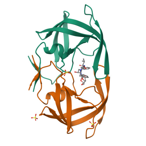
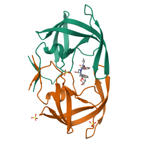
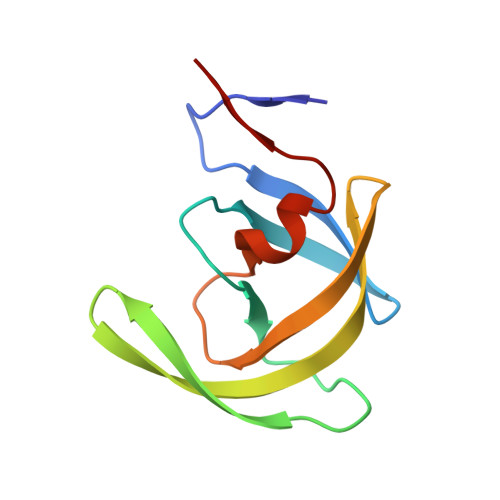
RCSB PDB - 6PJH: HIV-1 Protease NL4-3 WT in Complex with LR3-28

 6PJH

HIV-1 Protease NL4-3 WT in Complex with LR3-28


Experimental Data Snapshot

  • Method: X-RAY DIFFRACTION
  • Resolution: 1.85 Å
  • R-Value Free: 0.219 
  • R-Value Work: 0.185 
  • R-Value Observed: 0.187 

Starting Model: experimental
View more details

wwPDB Validation   3D Report Full Report


This is version 1.3 of the entry. See complete history


Literature

Structural Analysis of Potent Hybrid HIV-1 Protease Inhibitors Containing Bis-tetrahydrofuran in a Pseudosymmetric Dipeptide Isostere.

Rusere, L.N.Lockbaum, G.J.Henes, M.Lee, S.K.Spielvogel, E.Rao, D.N.Kosovrasti, K.Nalivaika, E.A.Swanstrom, R.Kurt Yilmaz, N.Schiffer, C.A.Ali, A.

(2020) J Med Chem 63: 8296-8313

  • DOI: https://doi.org/10.1021/acs.jmedchem.0c00529
  • Primary Citation of Related Structures:  
    6PJB, 6PJC, 6PJD, 6PJE, 6PJF, 6PJG, 6PJH, 6PJI, 6PJK, 6PJL, 6PJM, 6PJN, 6PJO

  • PubMed Abstract: 

    The design, synthesis, and X-ray structural analysis of hybrid HIV-1 protease inhibitors (PIs) containing bis-tetrahydrofuran (bis-THF) in a pseudo-C 2 -symmetric dipeptide isostere are described. A series of PIs were synthesized by incorporating bis-THF of darunavir on either side of the Phe-Phe isostere of lopinavir in combination with hydrophobic amino acids on the opposite P2/P2' position. Structure-activity relationship studies indicated that the bis-THF moiety can be attached at either the P2 or P2' position without significantly affecting potency. However, the group on the opposite P2/P2' position had a dramatic effect on potency depending on the size and shape of the side chain. Cocrystal structures of inhibitors with wild-type HIV-1 protease revealed that the bis-THF moiety retained similar interactions as observed in the darunavir-protease complex regardless of the position on the Phe-Phe isostere. Analyses of cocrystal structures and molecular dynamics simulations provide insights into optimizing HIV-1 PIs containing bis-THF in non-sulfonamide dipeptide isosteres.


  • Organizational Affiliation

    Department of Biochemistry and Molecular Pharmacology, University of Massachusetts Medical School, Worcester, Massachusetts 01605, United States.


Macromolecules
Find similar proteins by:  (by identity cutoff)  |  3D Structure
Entity ID: 1
MoleculeChains Sequence LengthOrganismDetailsImage
Protease NL4-3A [auth B],
B [auth A]
99Human immunodeficiency virus 1Mutation(s): 1 
Gene Names: pol
UniProt
Find proteins for P12497 (Human immunodeficiency virus type 1 group M subtype B (isolate NY5))
Explore P12497 
Go to UniProtKB:  P12497
Entity Groups  
Sequence Clusters30% Identity50% Identity70% Identity90% Identity95% Identity100% Identity
UniProt GroupP12497
Sequence Annotations
Expand
  • Reference Sequence
Small Molecules
Binding Affinity Annotations 
IDSourceBinding Affinity
OQG BindingDB:  6PJH Ki: min: 0.07, max: 0.3 (nM) from 3 assay(s)
Experimental Data & Validation

Experimental Data

  • Method: X-RAY DIFFRACTION
  • Resolution: 1.85 Å
  • R-Value Free: 0.219 
  • R-Value Work: 0.185 
  • R-Value Observed: 0.187 
  • Space Group: P 21 21 21
Unit Cell:
Length ( Å )Angle ( ˚ )
a = 51.546α = 90
b = 58.718β = 90
c = 61.991γ = 90
Software Package:
Software NamePurpose
HKL-2000data scaling
PHASERphasing
PHENIXrefinement
Cootmodel building

Structure Validation

View Full Validation Report


Entry History & Funding Information

Deposition Data


Funding OrganizationLocationGrant Number
National Institutes of Health/National Institute of General Medical Sciences (NIH/NIGMS)United StatesP01-GM109767

Revision History  (Full details and data files)

  • Version 1.0: 2020-07-01
    Type: Initial release
  • Version 1.1: 2020-07-29
    Changes: Database references
  • Version 1.2: 2020-08-26
    Changes: Database references
  • Version 1.3: 2023-10-11
    Changes: Data collection, Database references, Refinement description