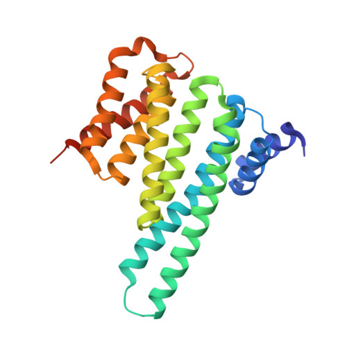
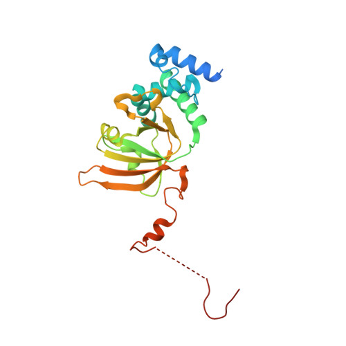
6GNJ

Exoenzyme S from Pseudomonas aeruginosa in complex with human 14-3-3 protein beta, trimeric crystal form in complex with STO1101


Experimental Data Snapshot

  • Method: X-RAY DIFFRACTION
  • Resolution: 3.24 Å
  • R-Value Free: 0.221 
  • R-Value Work: 0.173 
  • R-Value Observed: 0.177 

Starting Model: experimental
View more details

wwPDB Validation   3D Report Full Report


Ligand Structure Quality Assessment 


This is version 1.1 of the entry. See complete history


Literature

14-3-3 proteins activate Pseudomonas exotoxins-S and -T by chaperoning a hydrophobic surface.

Karlberg, T.Hornyak, P.Pinto, A.F.Milanova, S.Ebrahimi, M.Lindberg, M.Pullen, N.Nordstrom, A.Loverli, E.Caraballo, R.Wong, E.V.Nareoja, K.Thorsell, A.G.Elofsson, M.De La Cruz, E.M.Bjorkegren, C.Schuler, H.

(2018) Nat Commun 9: 3785-3785

  • DOI: https://doi.org/10.1038/s41467-018-06194-1
  • Primary Citation of Related Structures:  
    6GN0, 6GN8, 6GNJ, 6GNK, 6GNN

  • PubMed Abstract: 

    Pseudomonas are a common cause of hospital-acquired infections that may be lethal. ADP-ribosyltransferase activities of Pseudomonas exotoxin-S and -T depend on 14-3-3 proteins inside the host cell. By binding in the 14-3-3 phosphopeptide binding groove, an amphipathic C-terminal helix of ExoS and ExoT has been thought to be crucial for their activation. However, crystal structures of the 14-3-3β:ExoS and -ExoT complexes presented here reveal an extensive hydrophobic interface that is sufficient for complex formation and toxin activation. We show that C-terminally truncated ExoS ADP-ribosyltransferase domain lacking the amphipathic binding motif is active when co-expressed with 14-3-3. Moreover, swapping the amphipathic C-terminus with a fragment from Vibrio Vis toxin creates a 14-3-3 independent toxin that ADP-ribosylates known ExoS targets. Finally, we show that 14-3-3 stabilizes ExoS against thermal aggregation. Together, this indicates that 14-3-3 proteins activate exotoxin ADP-ribosyltransferase domains by chaperoning their hydrophobic surfaces independently of the amphipathic C-terminal segment.


  • Organizational Affiliation

    Department of Biosciences and Nutrition, Karolinska Institutet, Hälsovägen 4c, 14157, Huddinge, Sweden.


Macromolecules
Find similar proteins by:  (by identity cutoff)  |  3D Structure
Entity ID: 1
MoleculeChains Sequence LengthOrganismDetailsImage
14-3-3 protein beta/alpha
A, B
243Homo sapiensMutation(s): 0 
Gene Names: YWHAB
UniProt & NIH Common Fund Data Resources
Find proteins for P31946 (Homo sapiens)
Explore P31946 
Go to UniProtKB:  P31946
PHAROS:  P31946
GTEx:  ENSG00000166913 
Entity Groups  
Sequence Clusters30% Identity50% Identity70% Identity90% Identity95% Identity100% Identity
UniProt GroupP31946
Sequence Annotations
Expand
  • Reference Sequence
Find similar proteins by:  (by identity cutoff)  |  3D Structure
Entity ID: 2
MoleculeChains Sequence LengthOrganismDetailsImage
Exoenzyme S244Pseudomonas aeruginosaMutation(s): 2 
Gene Names: exoS
UniProt
Find proteins for Q93SQ1 (Pseudomonas aeruginosa)
Explore Q93SQ1 
Go to UniProtKB:  Q93SQ1
Entity Groups  
Sequence Clusters30% Identity50% Identity70% Identity90% Identity95% Identity100% Identity
UniProt GroupQ93SQ1
Sequence Annotations
Expand
  • Reference Sequence
Small Molecules
Ligands 1 Unique
IDChains Name / Formula / InChI Key2D Diagram3D Interactions
F4W
Query on F4W

Download Ideal Coordinates CCD File 
D [auth C]3-(12-oxidanylidene-7-thia-9,11-diazatricyclo[6.4.0.0^{2,6}]dodeca-1(8),2(6),9-trien-10-yl)propanoic acid
C12 H12 N2 O3 S
XKPJAYXICQQVTD-UHFFFAOYSA-N
Binding Affinity Annotations 
IDSourceBinding Affinity
F4W BindingDB:  6GNJ IC50: min: 6918, max: 1.90e+4 (nM) from 3 assay(s)
Experimental Data & Validation

Experimental Data

  • Method: X-RAY DIFFRACTION
  • Resolution: 3.24 Å
  • R-Value Free: 0.221 
  • R-Value Work: 0.173 
  • R-Value Observed: 0.177 
  • Space Group: C 1 2 1
Unit Cell:
Length ( Å )Angle ( ˚ )
a = 160.398α = 90
b = 59.357β = 125.84
c = 120.641γ = 90
Software Package:
Software NamePurpose
BUSTERrefinement
XDSdata reduction
SCALAdata scaling
PHASERphasing

Structure Validation

View Full Validation Report


Ligand Structure Quality Assessment 


Entry History & Funding Information

Deposition Data


Funding OrganizationLocationGrant Number
SwedenSB12-0022

Revision History  (Full details and data files)

  • Version 1.0: 2018-09-26
    Type: Initial release
  • Version 1.1: 2024-01-17
    Changes: Data collection, Database references, Refinement description