6GNN | pdb_00006gnn

Exoenzyme T from Pseudomonas aeruginosa in complex with human 14-3-3 protein beta, tetrameric crystal form bound to STO1101


Experimental Data Snapshot

  • Method: X-RAY DIFFRACTION
  • Resolution: 3.79 Å
  • R-Value Free: 
    0.385 (Depositor), 0.396 (DCC) 
  • R-Value Work: 
    0.285 (Depositor), 0.290 (DCC) 
  • R-Value Observed: 
    0.290 (Depositor) 

Starting Model: experimental
View more details

wwPDB Validation 3D Report Full Report

Validation slider image for 6GNN

Ligand Structure Quality Assessment 


This is version 1.1 of the entry. See complete history

Literature

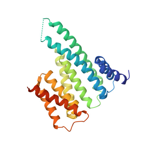
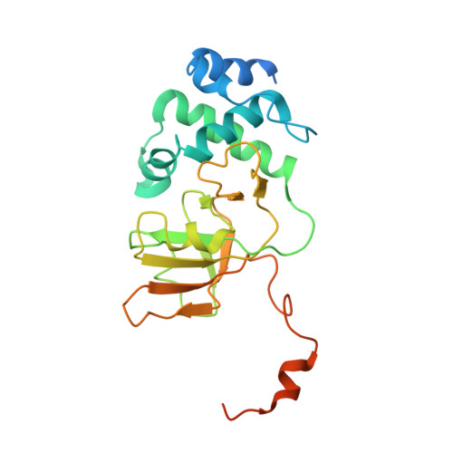
14-3-3 proteins activate Pseudomonas exotoxins-S and -T by chaperoning a hydrophobic surface.

Karlberg, T.Hornyak, P.Pinto, A.F.Milanova, S.Ebrahimi, M.Lindberg, M.Pullen, N.Nordstrom, A.Loverli, E.Caraballo, R.Wong, E.V.Nareoja, K.Thorsell, A.G.Elofsson, M.De La Cruz, E.M.Bjorkegren, C.Schuler, H.

(2018) Nat Commun 9: 3785-3785

  • DOI: https://doi.org/10.1038/s41467-018-06194-1
  • Primary Citation Related Structures: 
    6GN0, 6GN8, 6GNJ, 6GNK, 6GNN

  • PubMed Abstract: 

    Pseudomonas are a common cause of hospital-acquired infections that may be lethal. ADP-ribosyltransferase activities of Pseudomonas exotoxin-S and -T depend on 14-3-3 proteins inside the host cell. By binding in the 14-3-3 phosphopeptide binding groove, an amphipathic C-terminal helix of ExoS and ExoT has been thought to be crucial for their activation. However, crystal structures of the 14-3-3β:ExoS and -ExoT complexes presented here reveal an extensive hydrophobic interface that is sufficient for complex formation and toxin activation. We show that C-terminally truncated ExoS ADP-ribosyltransferase domain lacking the amphipathic binding motif is active when co-expressed with 14-3-3. Moreover, swapping the amphipathic C-terminus with a fragment from Vibrio Vis toxin creates a 14-3-3 independent toxin that ADP-ribosylates known ExoS targets. Finally, we show that 14-3-3 stabilizes ExoS against thermal aggregation. Together, this indicates that 14-3-3 proteins activate exotoxin ADP-ribosyltransferase domains by chaperoning their hydrophobic surfaces independently of the amphipathic C-terminal segment.


  • Organizational Affiliation
    • Department of Biosciences and Nutrition, Karolinska Institutet, Hälsovägen 4c, 14157, Huddinge, Sweden.

Macromolecule Content 

  • Total Structure Weight: 55.55 kDa 
  • Atom Count: 3,322 
  • Modeled Residue Count: 423 
  • Deposited Residue Count: 494 
  • Unique protein chains: 2

Macromolecules

Find similar proteins by:|  3D Structure
Entity ID: 1
MoleculeChains  Sequence LengthOrganismDetailsImage
14-3-3 protein beta/alpha248Homo sapiensMutation(s): 0 
Gene Names: YWHAB
UniProt & NIH Common Fund Data Resources
Find proteins for P31946 (Homo sapiens)
Explore P31946 
Go to UniProtKB:  P31946
PHAROS:  P31946
GTEx:  ENSG00000166913 
Entity Groups
Sequence Clusters30% Identity50% Identity70% Identity90% Identity95% Identity100% Identity
UniProt GroupP31946
Sequence Annotations
Expand
Reference Sequence
Find similar proteins by:|  3D Structure
Entity ID: 2
MoleculeChains  Sequence LengthOrganismDetailsImage
Exoenzyme TB [auth C]246Pseudomonas aeruginosa PAO1Mutation(s): 0 
Gene Names: exoTPA0044
EC: 2.4.2.31
UniProt
Find proteins for Q9I788 (Pseudomonas aeruginosa (strain ATCC 15692 / DSM 22644 / CIP 104116 / JCM 14847 / LMG 12228 / 1C / PRS 101 / PAO1))
Explore Q9I788 
Go to UniProtKB:  Q9I788
Entity Groups
Sequence Clusters30% Identity50% Identity70% Identity90% Identity95% Identity100% Identity
UniProt GroupQ9I788
Sequence Annotations
Expand
Reference Sequence

Small Molecules

Ligands 1 Unique
IDChains Name / Formula / InChI Key2D Diagram3D Interactions
F4W

Query on F4W


Download:Ideal Coordinates CCD File
C
3-(12-oxidanylidene-7-thia-9,11-diazatricyclo[6.4.0.0^{2,6}]dodeca-1(8),2(6),9-trien-10-yl)propanoic acid
C12 H12 N2 O3 S
XKPJAYXICQQVTD-UHFFFAOYSA-N

Experimental Data & Validation

Experimental Data

  • Method: X-RAY DIFFRACTION
  • Resolution: 3.79 Å
  • R-Value Free:  0.385 (Depositor), 0.396 (DCC) 
  • R-Value Work:  0.285 (Depositor), 0.290 (DCC) 
  • R-Value Observed: 0.290 (Depositor) 
Space Group: P 21 21 2
Unit Cell:
Length ( Å )Angle ( ˚ )
a = 115.378α = 90
b = 60.301β = 90
c = 81.19γ = 90
Software Package:
Software NamePurpose
PHENIXrefinement
DIALSdata reduction
DIALSdata scaling
PHASERphasing

Structure Validation

View Full Validation Report


Ligand Structure Quality Assessment 


Entry History 

& Funding Information

Deposition Data


Funding OrganizationLocationGrant Number
SwedenSB12-0022

Revision History  (Full details and data files)

  • Version 1.0: 2018-09-26
    Type: Initial release
  • Version 1.1: 2024-01-17
    Changes: Data collection, Database references, Refinement description